


Shopping Cart
Remove All Your shopping cart is currently empty
| Size | Quantity | Unit Price | Amount | Operation |
|---|
| Product |
|---|
| Bioactive |
| Species |
| Expression System |
| Tag |
| Accession Number |
| Amino Acid |
| Construction |
| Molecular Weight |
| Endotoxin |
| Formulation |
| Reconstitution |
| Stability & Storage |
| Shipping |
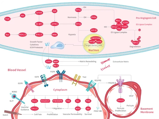
| Research Background |
| Size |
 Hello! How can I help you today?
 Copyright © 2015-2026 TargetMol Chemicals Inc. All Rights Reserved.