Shopping Cart
Remove All Your shopping cart is currently empty
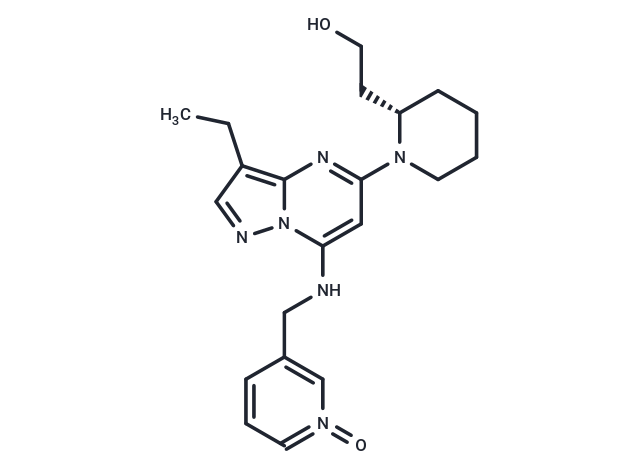
Dinaciclib (SCH 727965) is a selective CDK inhibitor targeting CDK1, CDK2, CDK5, and CDK9 (IC50 = 3/1/1/4 nM). It exhibits potential antitumor activity by inhibiting the incorporation of thoracic glycan (dThd) DNA.

| Pack Size | Price | USA Warehouse | Global Warehouse | Quantity |
|---|---|---|---|---|
| 2 mg | $47 | In Stock | In Stock | |
| 5 mg | $76 | In Stock | In Stock | |
| 10 mg | $121 | In Stock | In Stock | |
| 50 mg | $387 | In Stock | In Stock | |
| 1 mL x 10 mM (in DMSO) | $76 | In Stock | In Stock |
| Description | Dinaciclib (SCH 727965) is a selective CDK inhibitor targeting CDK1, CDK2, CDK5, and CDK9 (IC50 = 3/1/1/4 nM). It exhibits potential antitumor activity by inhibiting the incorporation of thoracic glycan (dThd) DNA. |
| Targets&IC50 | CDK9:4 nM (cell free), CDK5:1 nM (cell free), CDK2:1 nM (cell free), CDK1:3 nM (cell free) |
| In vitro | METHODS: Human pancreatic cancer cell lines MIAPaCa-2 and Pa20C were treated with Dinaciclib (0.5-128 nM) for 72 h. Cell viability was assayed using MTT Assay.
RESULTS: Dinaciclib inhibited the growth of MIAPaCa-2 and Pa20C cells in a dose-dependent manner, with a GI50 of approximately 10 and 20 nM, respectively.[1] METHODS: Human osteosarcoma cell lines SaOs-2 and U2OS were treated with Dinaciclib (5-62 nmol/L) for 4-24 h, and the expression levels of target proteins were detected using Western Blot. RESULTS: Dinaciclib eliminated the phosphorylation of CDK substrates in osteosarcoma cells. [2] |
| In vivo | METHODS: To assay antitumor activity in vivo, Dinaciclib (40 mg/kg, 20% (w/v) HPBCD) was injected intraperitoneally twice weekly for four weeks into CD1 nu/nu athymic mice harboring human pancreatic cancer tumors, JH033 or Panc286.
RESULTS: Dinaciclib inhibited the in vivo growth of a group of hypo-permeable cancer xenografts. [1] METHODS: To assay antitumor activity in vivo, Dinaciclib (8-48 mg/kg) was injected intraperitoneally into BALB/c mice harboring ovarian cancer tumor A2780 once daily for ten days. RESULTS: Dinaciclib had dose-dependent antitumor activity in vivo and almost completely inhibited tumor growth at dose levels below MTD. [3] |
| Kinase Assay | Recombinant cyclin/CDK holoenzymes were purified from Sf9 cells engineered to produce baculoviruses that express a specific cyclin or CDK. Cyclin/CDK complexes were typically diluted to a final concentration of 50 μg/mL in a kinase reaction buffer containing 50 mmol/L Tris-HCl (pH 8.0), 10 mmol/L MgCl2, 1 mmol/L DTT, and 0.1 mmol/L sodium orthovanadate. For each kinase reaction, 1 μg of enzyme and 20 μL of a 2-μmol/L substrate solution (a biotinylated peptide derived from histone H1) were mixed and combined with 10 μL of diluted SCH 727965. The reaction was started by the addition of 50 μL of 2 μmol/L ATP and 0.1 μCi of 33P-ATP. Kinase reactions were incubated for 1 hour at room temperature and were stopped by the addition of 0.1% Triton X-100, 1 mmol/L ATP, 5 mmol/L EDTA, and 5 mg/mL streptavidin-coated SPA beads. SPA beads were captured using a 96-well GF/B filter plate and a Filtermate universal harvester. Beads were washed twice with 2 mol/L NaCl and twice with 2 mol/L NaCl containing 1% phosphoric acid. The signal was then assayed using a TopCount 96-well liquid scintillation counter. Dose-response curves were generated from duplicate, eight-point serial dilutions of inhibitory compounds. IC50 values were derived by nonlinear regression analysis [1]. |
| Cell Research | A2780 cells were plated into six-well tissue culture dishes and allowed to adhere. Cells were then exposed to differing concentrations of SCH 727965 or a DMSO control vehicle for 24 hours, followed by a brief (30 min) pulsed exposure to bromodeoxyuridine (BrdUrd). Cells were then harvested, immunostained using FITC-conjugated antibodies specific for BrdUrd, counter-stained with propidium iodide/RNase A solution, and analyzed using flow cytometry. Fluorescence-activated cell sorting analyses were done on a FACSCalibur instrument. FITC-positive BrdUrd staining and propidium iodide signal allowed assessment of ongoing DNA replication and the cell cycle stage. Percentages of the cell population in each cell cycle stage were plotted for each test article concentration [1]. |
| Animal Research | For tumor implantation, specific cell lines were grown in vitro, washed once with PBS, and resuspended in 50% Matrigel in PBS to a final concentration of 4 × 10^7 to 5 × 10^7 cells per milliliter. Nude mice were injected with 0.1 mL of this suspension s.c. in the flank region. Tumor length (L), width (W), and height (H) were measured by a caliper twice weekly on each mouse and then used to calculate tumor volume using the formula (L × W × H)/2. When the tumor volume reached ~100 mm^3, the animals were randomized to treatment groups (10 mice/group) and treated i.p. with either SCH 727965 or individual chemotherapeutic agents according to the dosing schedule indicated in table and figure legends. Tumor volumes and body weights were measured during and after the treatment periods. Data were expressed as means ± SEM. Animals were euthanized according to the Institutional Animal Care and Use Committee guidelines [1]. |
| Synonyms | SCH 727965, PS-095760 |
| Molecular Weight | 396.49 |
| Formula | C21H28N6O2 |
| Cas No. | 779353-01-4 |
| Smiles | N(CC1=CN(=O)=CC=C1)C=2N3C(N=C(C2)N4[C@H](CCO)CCCC4)=C(CC)C=N3 |
| Relative Density. | 1.33 g/cm3 |
| Color | Yellow |
| Appearance | Solid |
| Storage | Powder: -20°C for 3 years | In solvent: -80°C for 1 year | Shipping with blue ice/Shipping at ambient temperature. | ||||||||||||||||||||||||||||||||||||||||
| Solubility Information | DMSO: 130 mg/mL (327.88 mM), Sonication is recommended. Ethanol: 8 mg/mL (20.18 mM), Heating is recommended. | ||||||||||||||||||||||||||||||||||||||||
| In Vivo Formulation | 10% DMSO+40% PEG300+5% Tween 80+45% Saline: 5 mg/mL (12.61 mM), Solution. Please add the solvents sequentially, clarifying the solution as much as possible before adding the next one. Dissolve by heating and/or sonication if necessary. Working solution is recommended to be prepared and used immediately. The formulation provided above is for reference purposes only. In vivo formulations may vary and should be modified based on specific experimental conditions. | ||||||||||||||||||||||||||||||||||||||||
Solution Preparation Table | |||||||||||||||||||||||||||||||||||||||||
Ethanol/DMSO
DMSO
| |||||||||||||||||||||||||||||||||||||||||
| Size | Quantity | Unit Price | Amount | Operation |
|---|

Copyright © 2015-2026 TargetMol Chemicals Inc. All Rights Reserved.