


Shopping Cart
Remove All Your shopping cart is currently empty
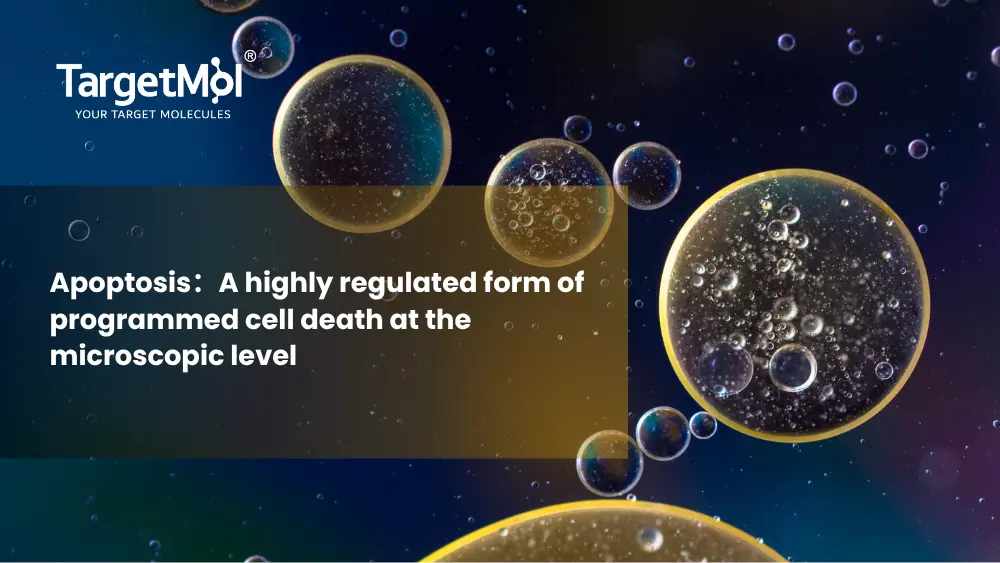
In life science research, apoptosis has become an indispensable experimental tool and a critical biological process of interest. It is extensively studied in tumor biology, where dysregulation of apoptotic pathways contributes to uncontrolled cell proliferation and cancer progression. In neurodegenerative diseases such as Alzheimer’s and Parkinson’s disease, excessive neuronal apoptosis is closely associated with disease pathogenesis and progression. Apoptosis research is also highly relevant in autoimmune disorders, where abnormal survival or elimination of immune cells can disrupt immune tolerance and lead to chronic inflammation.
Core Mechanisms of Apoptosis
Apoptosis is tightly regulated by genes, giving it high selectivity and distinct morphological characteristics. It generally consists of three stages: triggering signals, signal transduction, and execution.
• Triggering signals:
These are usually intracellular or extracellular stress factors, such as DNA damage, oxidative stress, or the binding of death receptors and ligands (e.g., Fas/FasL). Once the apoptotic signal persists or reaches a critical threshold, the cell irreversibly proceeds toward death.
• Signal transduction:
Key mediators include the Bcl-2 family proteins, mitochondrial release of cytochrome c, and the death-inducing signaling complex (DISC), which amplify apoptotic signals through cascade pathways.
• Execution phase:
Members of the Caspase family (such as Caspase-3, Caspase-8, and Caspase-9) specifically cleave critical structural and functional proteins within the cell, ultimately leading to cellular disassembly.
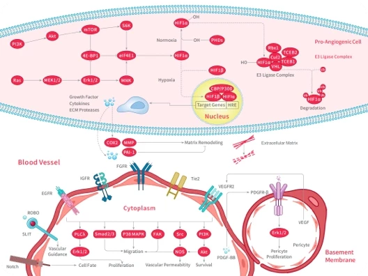
Major Apoptotic Pathways
According to different triggering mechanisms, apoptosis can mainly be classified into the extrinsic pathway (death receptor pathway), the intrinsic pathway (mitochondrial pathway), and the endoplasmic reticulum stress pathway.
Depending on the research context, apoptosis studies can generally be divided into:
Pro-apoptotic research in tumor cells, aimed at inducing cancer cell death; and
Anti-apoptotic protective research in normal cells (such as neurons and cardiomyocytes), focused on preventing excessive cell death and tissue damage.
Common Research Targets in Apoptosis
✅ Bcl-2 Family:
The “gatekeepers” of the mitochondrial pathway. The balance between anti-apoptotic proteins (such as Bcl-2/Bcl-xL) and pro-apoptotic proteins (such as Bax/Bak) determines cell fate, making them high-frequency targets in the development of therapies for hematologic malignancies and other cancers.
✅ Caspase Family:
The “executioners” of apoptosis. Initiator Caspases and effector Caspases amplify apoptotic signaling through cascade activation. Their activation marks the irreversible stage of apoptosis and is considered the gold standard for evaluating apoptotic activity.
✅ p53 Tumor Suppressor Protein:
Upon DNA damage, p53 is activated to upregulate pro-apoptotic gene expression, inducing cell cycle arrest or apoptosis.
✅ Death Receptors:
These receptors trigger the extrinsic apoptotic pathway through binding with corresponding ligands (such as FasL and TNF-α). They play critical roles in immune regulation and anti-tumor immunity.
✅ IAPs (Inhibitor of Apoptosis Proteins):
By surface modification or direct binding, IAPs can suppress Caspase activity. They maintain cellular homeostasis by preventing excessive apoptosis and are widely studied in tumor drug resistance research.
How to Choose Apoptosis Modulators with Different Mechanisms of Action?
The selection of apoptosis modulators is highly dependent on your research objectives. When choosing an appropriate modulator, both target specificity and experimental compatibility should be carefully considered:
|
Mechanism |
Apoptosis Inducers |
Apoptosis Inhibitors |
|
Advantages |
- High targeting specificity, capable of selectively activating specific apoptotic pathways - Rapid response - Effectively eliminates abnormally proliferating cells |
- Potently blocks apoptotic cascade reactions (e.g., pan-Caspase inhibition) - Structurally stable with long-lasting activity - Protects vulnerable cells |
|
Application |
- Anttumor drug screening - Drug resistance mechanism analysis - Establishment of positive control models for apoptosis studies |
- Neurodegenerative disease models - Cardiovascular ischemia-reperfusion injury - Validation of whether specific phenotypes are dependent on apoptotic pathways |
What Is the Standard Workflow for Apoptosis Detection?
In general, cells are first seeded into plates and allowed to reach an appropriate density before use. Apoptosis inducers or target compounds are then added, and the cells are incubated for a certain period (typically 12–48 hours) to trigger the apoptotic process.
After treatment, the cells are washed to remove nonspecific substances, followed by staining with fluorescent probes, such as the Annexin V-FITC/PI dual-staining method, for subsequent analysis by flow cytometry or fluorescence microscopy.
Throughout the experiment, both positive and negative controls should be included, and mechanical damage during cell collection should be minimized to avoid false-positive results.
Related Products
|
TSID |
Product |
|
T7020 |
Z-VAD(OH)-FMK (Irreversible broad-spectrum Caspase inhibitor that can be directly used in culture media without esterase pretreatment) |
|
T6013 |
Z-VAD(OMe)-FMK (Cell-permeable irreversible pan-Caspase inhibitor) |
|
T6680 |
Staurosporine (Broad-spectrum kinase inhibitor / Classic positive control for apoptosis induction) |
|
T7019 |
Z-IETD-FMK (A cell-permeable selective Caspase-8 inhibitor) |
|
T6005 |
Z-DEVD-FMK (A cell-permeable selective Caspase-3 inhibitor) |
|
T2503 |
PAC-1 (Procaspase-3 activator, commonly used in studies of inducing apoptosis in cancer cells) |
|
L9000 |
Apoptosis Compound Library (Apoptosis Compound Library containing 2,000+ bioactive small molecules targeting apoptosis pathways) |
An essential round-up of science news, opinion and analysis, delivered to your inbox every weekday.
 Hello! How can I help you today?
 Copyright © 2015-2026 TargetMol Chemicals Inc. All Rights Reserved.