


Shopping Cart
Remove All Your shopping cart is currently empty
TargetMol Star Product—MitoSOX Red (T75342, CAS1003197-00-9), a fluorescent tool for visualizing mitochondrial redox status
MitoSOX Red (Cat. No. T75342, CAS No. 1003197-00-9) is a novel fluorescent probe that is specific, cell-permeable, and targets mitochondria in living cells.
Upon entering mitochondria, MitoSOX Red is oxidized by superoxide but not by other ROS or RNS-generating systems. MitoSOX Red then binds to mitochondrial nucleic acids, producing intense red fluorescence. MitoSOX Red serves as a fluorescent indicator for the specific detection of superoxide. Superoxide dismutase (SOD) can prevent the oxidation of MitoSOX Red.
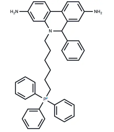
Molecular Structure of MitoSOX Red
1. Background
A novel fluorescent probe targeting mitochondria in living cells, this tool represents a cutting-edge advancement in the fields of cell biology and biomedical imaging. Its core innovations include long-term stable labeling, super-resolution compatibility, near-infrared/two-photon excitation, and multifunctional responses, enabling precise visualization of mitochondrial dynamics, microenvironments, and pathological changes.
The mitochondrial membrane potential (Δψm ≈ -180 mV, negative inside and positive outside) is the primary driving force enabling fluorescent probes to precisely “anchor” themselves to mitochondria. Their targeting mechanisms primarily revolve around group characteristics and novel strategies: Probe molecules typically carry cationic targeting groups, such as triphenylphosphonium (TPP⁺), pyridinium, and quinoline derivatives. These positively charged groups are specifically captured by the negative potential of the mitochondrial inner membrane and accumulate within the membrane. simultaneously,probes often possess lipophilic backbones such as cyanine (Cy), rhodamine, and BODIPY, which facilitate the probe’s smooth penetration of the cell membrane and the outer mitochondrial membrane, thereby achieving initial targeting of mitochondria.
MitoSOX Red utilizes a TPP⁺ targeting module and an oxidation-sensitive dihydroethidine core to achieve precise enrichment and specific detection of superoxide (O₂・⁻) in live-cell mitochondria. Driven by the negative mitochondrial membrane potential, the system prevents off-target binding to other organelles. Upon oxidation by superoxide, it generates strong red fluorescence that is not interfered with by other ROS/RNS. SOD can inhibit its oxidation, ensuring quantitative accuracy. Compatible with multiple platforms including fluorescence microscopy and flow cytometry, it supports long-term dynamic monitoring and is widely used in disease mechanism studies, drug screening, and toxicological assessments. With its high specificity, high precision, and live-cell compatibility, it has become a key tool in mitochondrial biology research, driving breakthroughs in oxidative stress-related fields.
2. Selected Application Literature
2.1 Article Title:Herceptin induces ferroptosis and mitochondrial dysfunction in H9c2cells
Study Overview: Using rat H9c2 cardiomyocytes as a model, this study demonstrated that Herceptin (trastuzumab) causes cardiomyocyte damage by inducing ferroptosis and mitochondrial dysfunction, which constitutes a key mechanism underlying its clinical cardiotoxicity. The ferroptosis inhibitor Ferrostatin-1 (Fer-1) and the iron chelator Deferoxamine (DFO) can significantly reverse the aforementioned damage, providing new therapeutic targets for the prevention and treatment of Herceptin-associated cardiomyopathy. [2]
MitoSOX Red served as the core probe in this study for the specific detection of mitochondrial superoxide in live cells, used to elucidate the mechanism of Herceptin-induced myocardial damage. It precisely tracks ROS of mitochondrial origin, demonstrating that Herceptin induces mitochondrial oxidative stress in a dose- and time-dependent manner. it also validates that Fer-1/DFO exerts a protective effect by reversing mitochondrial superoxide bursts. These results are corroborated by measurements of mitochondrial membrane potential, ATP levels, and ferroptosis markers, fully elucidating the pathway: “elevated mitochondrial ROS → oxidative stress → ferroptosis → myocardial injury.”
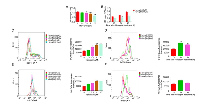
MitoSOX Red revealed a significant increase in Herceptin-induced mitochondrial superoxide levels.
2.2 Article Title:Mitochondrial DNA leakage induces odontoblast inflammation via the cGAS-STING pathway

Research Overview: This study demonstrates that LPS stimulation leads to mitochondrial damage in odontoblasts → leakage of mtDNA into the cytoplasm → activation of the cGAS-STING pathway → promotion of the release of inflammatory factors such as IL-6 and CXCL10, ultimately triggering pulpitis. MitoSOX Red, used for the specific detection of mitochondrial superoxide, serves as a key tool for demonstrating the sequence of mitochondrial oxidative stress → mitochondrial damage → mtDNA leakage. The mtDNA-cGAS-STING axis can serve as a new therapeutic target for pulpitis. [3]
MitoSOX Red specifically detects mitochondrial superoxide anions, directly demonstrating that LPS induces mitochondrial oxidative stress—a critical upstream event leading to mitochondrial structural damage and mtDNA leakage—and provides direct fluorescent evidence for the mtROS → mtDNA leakage → cGAS-STING → inflammation pathway.
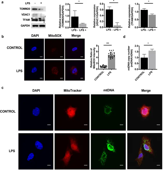
MitoSOX Red indicates a significant increase in mitochondrial superoxide
2.3 Article Title:Ferrostatin-1 ameliorates Cis-dichlorodiammineplatinum(II)-induced ovarian toxicity by inhibiting ferroptosis
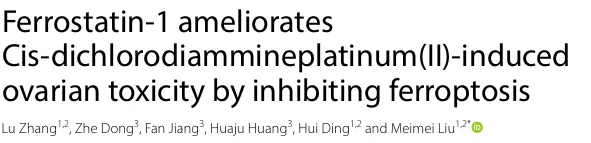
Study Overview: This study demonstrates that cisplatin (CDDP) induces ferroptosis by causing iron overload in ovarian granulosa cells, a burst of mitochondrial ROS, mitochondrial dysfunction, and lipid peroxidation, leading to ovarian damage and premature ovarian failure (POF). Ferrostatin-1 (FER-1) can inhibit ferroptosis by targeting the GPX4/NRF2 pathway, significantly alleviating CDDP-induced ovarian toxicity and restoring hormone levels and ovarian function. MitoSOX Red-Red, as a specific probe for mitochondrial superoxide, serves as a key tool for demonstrating that mitochondrial oxidative stress drives ferroptosis. [4]
This study utilized rat granulosa cells and a premature ovarian failure model to confirm that cisplatin (CDDP) induces ferroptosis through iron overload, oxidative stress, and mitochondrial damage, resulting in ovarian toxicity. Specific detection with MitoSOX Red-Red revealed that CDDP significantly increased mitochondrial superoxide levels, triggering oxidative stress and a decrease in membrane potential. Ferrostatin-1 (FER-1) could reverse these changes. Concurrently, FER-1 restored cell viability, improved mitochondrial morphology, and regulated the NRF2/HO-1/GPX4 pathway. it also exerted a protective effect by directly binding to GPX4 and NRF2, thereby providing a new strategy for alleviating chemotherapy-induced ovarian damage.
MitoSOX Red-Red specifically labels mitochondrial superoxide, validating the protective effect of FER-1
References
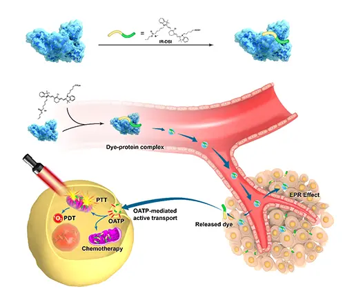
[1]Tan X,Luo S,Long L,et al.Structure-Guided Design and Synthesis of aMitochondria-Targeting Near-Infrared Fluorophore with Multimodal Therapeutic Activities.Adv Mater.2017.29(43):10.1002/adma.201704196. doi:10.1002/adma.201704196
[2] Sun L, Wang H, Yu S, Zhang L, Jiang J, Zhou Q. Herceptin induces ferroptosis and mitochondrial dysfunction in H9c2 cells. Int J Mol Med. 2022.49(2):17. doi:10.3892/ijmm.2021.5072
[3]Zhou L,Zhang YF,Yang FH,Mao HQ,Chen Z,Zhang L.Mitochondrial DNA leakage induces odontoblast inflammation via the cGAS-STING pathway.Cell Commun Signal.2021.19(1):58.Published2021May20.doi:10.1186/s12964-021-00738-7
[4]Zhang L,Dong Z,Jiang F,Huang H,Ding H,Liu M.Ferrostatin-1ameliorates Cis-dichlorodiammineplatinum(II)-induced ovarian toxicity by inhibiting ferroptosis.Mol Med.2024.30(1):150.Published2024Sep13.doi:10.1186/s10020-024-00923-7
An essential round-up of science news, opinion and analysis, delivered to your inbox every weekday.
 Hello! How can I help you today?
 Copyright © 2015-2026 TargetMol Chemicals Inc. All Rights Reserved.