


Shopping Cart
Remove All Your shopping cart is currently empty
TargetMol—Natural Product—3-Methyladenine (Cat. No. T1879, CAS. 5142-23-4), Switches that regulate autophagy flux
1. Product Introduction
3-Methyladenine (Cat. No. T1879, CAS. 5142-23-4), also known as NSC 66389, 3-MA.3-Methyladenine is a PI3K inhibitor that selectively inhibits IB-type PI3Kγ (IC50 = 60 μM) and III-type VPS34 (IC50 = 25 μM). 3-Methyladenine has autophagic inhibitory activity.
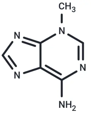
Molecular structure of 3-Methyladenine
2. Background Introduction
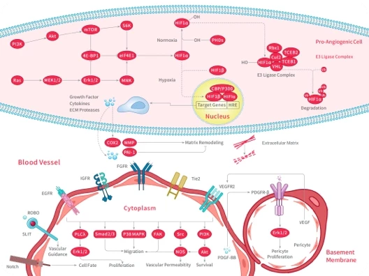
Autophagy is a highly conserved degradation pathway in cells. It is an important mechanism to maintain cell homeostasis and cope with stress by forming autophagosomes and fusing with lysosomes to achieve the degradation and reuse of damaged organelles, proteins and intracellular macromolecules. This process includes several steps, such as diaphragm initiation, extension and closure, formation and maturation of autophagosomes, and fusion with lysosomes. It is regulated by a series of proteins encoded by autophagy-related genes ( ATG ). In mammalian cells, autophagy is regulated by nutrition, energy and stress signals. Signaling pathways such as AMPK and mTORC1 become core regulatory nodes. The classical macroautophagy pathway initiates the process of organelle and macromolecular encapsulation to form autophagosomes. Autophagy plays a key role in a variety of physiological and pathological processes, such as cell survival, immune response, neurodegenerative diseases, tumor development, etc. Therefore, related molecular targets have become potential therapeutic intervention points. As an important enzyme in the initial stage of autophagy, class III PI3K ( Vps34 ) in the PI3K family is involved in the formation of phosphatidylinositol 3-phosphate, thus driving the formation of phagophore, which is one of the important regulatory targets of autophagy. [1]
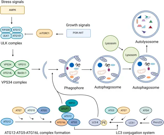
Flow chart of autophagy occurrence [1]
3-Methyladenine ( 3-MA ) is a classical autophagy inhibitor, which mainly inhibits autophagy by targeting class III PI3K ( Vps34 ) in the PI3K family. 3-Methyladenine can inhibit the lipid kinase activity of Vps34 and block the synthesis of PI ( 3 ) P, thereby interfering with the formation of autophagic membrane and the initial stage of autophagosome, resulting in a decrease in autophagy flux. In addition, 3-Methyladenine also has a certain inhibitory activity against class I PI3K.This dual targeting property makes it a common tool compound in the study of the interaction between PI3K / Akt / mTOR signaling axis and autophagy. By inhibiting Vps34-mediated autophagy initiation, 3-Methyladenine is often used in vitro and in vivo models to explore the role of autophagy in cell survival, metabolic regulation, and tumor drug sensitivity, and as a chemical intervention to verify autophagy-dependent phenotypes. [2]
3. Application References
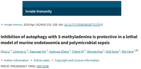
Inhibition of autophagy with 3-methyladenine is protective in a lethal model of murine endotoxemia and polymicrobial sepsis
Research Overview:
In this study, we established two lethal systemic inflammatory models in mice-LPS-induced endotoxemia model and cecal ligation and puncture ( CLP ) -induced multibacillary sepsis model, and systematically evaluated the effects of classical autophagy inhibitor 3-Methyladenine ( 3-MA ) on disease progression and survival outcomes. The study found that the level of autophagy was significantly up-regulated in these two inflammatory shock models. After inhibiting autophagy by intraperitoneal injection of 3-Methyladenine, the survival rate of mice was significantly improved, accompanied by decreased levels of inflammatory factors, reduced organ damage and improved histopathology. Mechanistically, the authors confirmed that 3-Methyladenine attenuates excessive inflammatory response and cell damage by inhibiting the Vps34-dependent autophagy initiation process, suggesting that autophagy is not a simple protective mechanism during severe infection and sepsis. Excessive activation may aggravate pathological damage. This study systematically confirmed the protective effect of autophagy inhibition in a lethal sepsis model for the first time in vivo, and provided an important experimental basis for understanding the dual role of autophagy in inflammatory diseases and its potential therapeutic targets. [3]
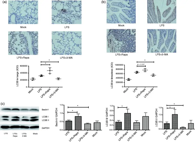
In endotoxic shock mice, LPS combined with autophagy inhibitor 3-Methyladenine or LPS combined with rapamycin inhibited or enhanced autophagy, respectively [3]
Modulation of O-GlcNAc cycling influences α-synuclein amplification, degradation, and associated neuroinflammatory pathology
Research Overview:
This study systematically explored the effects of regulating O-GlcNAc glycosylation cycle ( mediated by O-GlcNAc transferase OGT and O-GlcNAcase OGA ) on the aggregation, diffusion, degradation of pathological α-synuclein and related neuroinflammatory responses in cell and mouse models. Researchers injected α-syn prefibrils with different conformations into the mouse striatum or nerve / microglia co-culture system, and combined pharmacological or genetic methods to inhibit OGT or OGA activity. It was found that the regulation of O-GlcNAcylation level significantly changed the aggregation degree of α-syn and its transmission to adjacent cells and connected brain regions, and also affected the degeneration of dopaminergic neurons, NLRP3-mediated neuroinflammatory response and behavioral defects. In this process, pathological α-syn transmission reduces the level of O-GlcNAc and the expression of OGT in recipient cells, while inhibition of OGA to increase O-GlcNAcylation alleviates α-syn-related pathology and promotes its degradation through the autophagy-lysosomal pathway. [4]
In this study, 3-Methyladenine ( Cat. No. T1879 ) was used as a classical autophagy inhibitor to verify whether the clearance of α-synuclein is dependent on the autophagy-lysosomal pathway. The authors added 3-Methyladenine to the cell model by pharmacological means to inhibit the Vps34-dependent autophagy initiation process, and then observed the degradation of α-syn aggregates.
4. References
[1] Liu Y, Yang Q, Chen S, Li Z, Fu L. Targeting VPS34 in autophagy: An update on pharmacological small-molecule compounds. Eur J Med Chem. 2023 Aug 5;256:115467. doi: 10.1016/j.ejmech.2023.115467
[2] Klionsky DJ, Abdelmohsen K, Abe A, et al. Guidelines for the use and interpretation of assays for monitoring autophagy (3rd edition). Autophagy. 2016;12(1):1-222. doi: 10.1080/15548627.2015.1100356
[3] Li Q, Li L, Fei X, Zhang Y, Qi C, Hua S, Gong F, Fang M. Inhibition of autophagy with 3-methyladenine is protective in a lethal model of murine endotoxemia and polymicrobial sepsis. Innate Immun. 2018 May;24(4):231-239. doi: 10.1177/1753425918771170. Epub 2018 Apr 19. PMID: 29673286; PMCID: PMC6830927.
[4] Miao Y, Zhang T, Ma Z, Du H, Gu Q, Jiang M, Xiong K, Liu CF, Meng H. Modulation of O-GlcNAc cycling influences α-synuclein amplification, degradation, and associated neuroinflammatory pathology. Mol Neurodegener. 2025 Oct 27;20(1):113. doi: 10.1186/s13024-025-00904-2

An essential round-up of science news, opinion and analysis, delivered to your inbox every weekday.
 Hello! How can I help you today?
 Copyright © 2015-2026 TargetMol Chemicals Inc. All Rights Reserved.