
Shopping Cart
Remove All Your shopping cart is currently empty


Synonyms: RU486, RU 38486, C-1073
| Pack Size | Price | USA Stock | Global Stock | Quantity |
|---|---|---|---|---|
| 50 mg | $33 | In Stock | In Stock | |
| 100 mg | $48 | In Stock | In Stock | |
| 500 mg | $84 | In Stock | In Stock | |
| 1 g | $119 | In Stock | In Stock | |
| 1 mL x 10 mM (in DMSO) | $35 | In Stock | In Stock |
| Description | Mifepristone (C-1073) is a progesterone-receptor (IC50=0.2 nM) and glucocorticoid-receptor antagonist (IC50=2.6 nM). Mifepristone is used to terminate pregnancy, treat uterine fibroids, and treat endometriosis. |
| Targets & IC50 | T47D cells:0.045 nM, COS-7 cells:0.6 nM, MCF-7 cells:24.03 μM, SUM149PT cells:15 μM, Progesterone receptor:0.2 nM, HCC1937 cells:> 20 μM, CHO cells-K1 cells:0.4 nM, CHO cells:5 nM, LNCaP cells:11.9 nM (EC50), A549 cells:1.6 nM, GR:2.6 nM, K562 cells/R7:0.9 μM, HEK293 cells:0.298 nM |
| In vitro | METHODS: Human MCF7 cells were treated with Mifepristone (1-100 μM) for 72 hours, and the growth inhibition of the cells was detected by the CCK-8 method. RESULTS: Mifepristone inhibited the growth of MCF7 cells (IC50=24.03 μM). [1] METHODS: Ovarian cancer cells SK-OV-3 and OV2008 were treated with Mifepristone (5, 10, 15, 20 μM) for 3 days, and the number of surviving cells was evaluated using trypan blue staining elimination (Sigma). RESULTS: Mifepristone dose-dependently inhibited the growth of SK-OV-3 cells (IC50=6.25 μM) and OV2008 cells (IC50=6.91 μM). [2] |
| In vivo | METHODS: To study the anti-tumor activity of Mifepristone, Mifepristone (2 mg/kg) was subcutaneously injected into a nude mouse model of cervical tumor xenotransplantation for 3 consecutive days. Then Cisplatin (3 mg/kg) was intraperitoneally injected into nude mice for 3 consecutive days. RESULTS: Tumor growth was inhibited when treated with Cisplatin alone. The combination of Cisplatin and Mifepristone led to a more significant reduction in tumor weight, reducing it by approximately 50%. [3] METHODS: To study the anti-tumor activity of Mifepristone, Mifepristone (0.5, 1 mg/kg) was subcutaneously implanted into the subcutaneous graft tumor model established in nude mice by the SK-OV-3 ovarian cancer cell line. RESULTS: Mifepristone significantly inhibited tumor growth in a dose-dependent manner, and the effect was observed 20 days after the start of treatment. [2] METHODS: To study the anti-tumor activity of Mifepristone, Mifepristone (50 mg/kg) was subcutaneously injected into the subcutaneous graft tumor model established in nude mice by the MKN-45 gastric cancer cell line. RESULTS: Mifepristone significantly reduced the number of lung metastases. In transplanted tumors, Mifepristone downregulated the expressions of vascular endothelial growth factor (VEGF) and microvessel density (MVD). [4] METHODS: To study the anti-tumor activity of Mifepristone, Mifepristone (15 mg/kg) was subcutaneously injected into the subcutaneous transplanted tumor model established by the SK-N-SH neuroblastoma cell line in nude mice twice a week for a total of six times. RESULTS: Mifepristone significantly inhibited tumor growth, with an inhibition rate as high as 80%. The volume and weight of the tumor were significantly reduced after Mifepristone treatment. [5] |
| Synonyms | RU486, RU 38486, C-1073 |
| Kinase Assay | Glucocorticoid receptor (GR) antagonist activity, Progesterone receptor (PR) antagonist activity: T47D alkaline phosphatase assay: T47D human breast cancer cells are plated in 96-well tissue culture plates at 104 cells per well in assay medium [RPMI medium without phenol red containing 5% (v/v) charcoal-treated FBS and 1% (v/v) penicillin–streptomycin]. Two days later, the medium is decanted and Mifepristone or control is added at a final concentration of 0.1% (v/v) dimethylsulfoxide in fresh assay medium. Twenty-four hours later, an alkaline phosphatase assay is performed using a SEAP kit. The medium is decanted and the cells are fixed for 30 minutes at room temperature with 5% (v/v) formalin. The cells are washed once at room temperature with Hanks' buffered saline solution. Equal volumes (0.05 mL) of dilution buffer, assay buffer, and 1:20 substrate/enhancer mixture are then added. After 1-hour incubation in the dark at room temperature, the lysate is transferred to a white 96-well plate and luminescence is read using a LuminoSkan Ascen. A549 reporter assay: A549 human lung carcinoma cells are washed with OPTI-MEM I. The medium is removed and lipid–DNA complex solution (1.5 μg/mL of GRE-luciferase reporter DNA in 8.5 mL OPTI-MEM I plus 6 μL/mL DMRIE-C reagent in 8.5 mL OPTI-MEM I, combined, mixed and incubated at room temperature for 40 minutes) is overlayed onto the cells in a T160 flask. The cells are incubated for 16 hours at 37 °C in a CO2 incubator. The DNA-containing medium is removed and 30 mL of growth medium containing 5% (v/v) charcoal-treated fetal bovine serum is added. After 5-6 hours, the cells are seeded in 96-well plates and incubated overnight at 37 °C. Mifepristone is then added to each well followed by dexamethasone as a corticoid challenge. The cells are incubated for 24 hours. Luciferase assay buffer is added to each well and the cells are incubated for 30 minutes at room temperature.Luciferase activity is measured in a DYNEX Microlite plate on a TopCount. |
| Cell Research | Cell growth is evaluated in various ovarian cancer cell lines that are subjected to dose-response or time course treatments. Media containing each of the doses of fresh steroids is replaced every 24 hours. Control groups of cells are treated with vehicle ethanol at a final concentration of less than 0.05%. Number of viable cells is evaluated by trypsinization and counting in a hemocytometer chamber using trypan blue dye exclusion. Experiments are conducted in media without phenol red and supplemented with charcoal extracted fetal bovine serum, or media containing unextracted serum and having phenol red. Similar results are obtained with both media preparations; therefore, after performing the growth curves, all subsequent experiments are conducted using media with unextracted serum and in the presence of phenol red. When indicated, the proliferation IC50s are calculated using software designed to study drug interaction. (Only for Reference) |
| Molecular Weight | 429.59 |
| Formula | C29H35NO2 |
| Cas No. | 84371-65-3 |
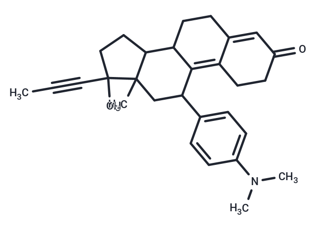
| Smiles | CC#CC1(O)CCC2C3CCC4=CC(=O)CCC4=C3C(CC12C)C1=CC=C(C=C1)N(C)C |
| Relative Density. | 1.18 g/cm3 |
| Storage | Powder: -20°C for 3 years | In solvent: -80°C for 1 year Shipping with blue ice/Shipping at ambient temperature. | ||||||||||||||||||||||||||||||||||||||||
| Solubility Information | DMSO: 255 mg/mL (593.59 mM), Sonication is recommended. Ethanol: 21.5 mg/mL (50.05 mM), Heating is recommended. | ||||||||||||||||||||||||||||||||||||||||
| In Vivo Formulation | 10% DMSO+40% PEG300+5% Tween 80+45% Saline: 4.3 mg/mL (10.01 mM), Suspension. Please add the solvents sequentially, clarifying the solution as much as possible before adding the next one. Dissolve by heating and/or sonication if necessary. Working solution is recommended to be prepared and used immediately. The formulation provided above is for reference purposes only. In vivo formulations may vary and should be modified based on specific experimental conditions. | ||||||||||||||||||||||||||||||||||||||||
Solution Preparation Table | |||||||||||||||||||||||||||||||||||||||||
Ethanol/DMSO
DMSO
Note : The dilution table applies only to solid products. For liquid products, please calculate the stock solution based on the stated concentration and/or density. | |||||||||||||||||||||||||||||||||||||||||
 For example, if the intended dosage is 10 mg/kg for animals weighing 20 g , with a dosing volume of 100 μL per animal, and a total of 10 animals are to be administered, using a formulation of
  10% DMSO+ 40% PEG300+ 5% Tween 80+ 45% Saline/PBS/ddH2O , the resulting working solution concentration would be 2 mg/mL.
Dissolve 2 mg of the compound in 100 μL DMSO to obtain a stock solution at a concentration of 20 mg/mL . If the required concentration exceeds the compound's known solubility, please contact us for technical support before proceeding.
1) Add 100 μL of the DMSO stock solution to 400 µL PEG300 and mix thoroughly until the solution becomes clear.
2) Add 50 µL Tween 80 and mix well until fully clarified.
3) Add 450 µL Saline,PBS or ddH2O and mix thoroughly until a homogeneous solution is obtained.
| Size | Quantity | Unit Price | Amount | Operation |
|---|
 Hello! How can I help you today?
 Copyright © 2015-2026 TargetMol Chemicals Inc. All Rights Reserved.
