
Shopping Cart
Remove All Your shopping cart is currently empty

Synonyms: nuclear receptor subfamily 3, group C, member 1 (glucocorticoid receptor), GRL, GR, GCRST, GCR, GCCR
Anti-NR3C1 Polyclonal Antibody 2
| Pack Size | Price | USA Stock | Global Stock | Quantity |
|---|---|---|---|---|
| 50 µL | $220 | 7-10 days | 7-10 days | |
| 100 µL | $373 | 7-10 days | 7-10 days | |
| 200 µL | $529 | 7-10 days | 7-10 days |
| Description | Anti-NR3C1 Polyclonal Antibody 2 is a Rabbit antibody targeting NR3C1. Anti-NR3C1 Polyclonal Antibody 2 can be used in FCM, IF, IHC-Fr, IHC-P, WB. |
| Synonyms | nuclear receptor subfamily 3, group C, member 1 (glucocorticoid receptor), GRL, GR, GCRST, GCR, GCCR |
| Ig Type | IgG |
| Reactivity | Human,Mouse,Rat (predicted:Chicken,Dog,Pig,Cow,Horse,Sheep,GuineaPig) |
| Verified Activity | 1. Blank control: A549. Primary Antibody (green line): Rabbit Anti-Glucocorticoid Receptor antibody (TMAB-01267) Dilution: 1 μg/10^6 cells; Isotype Control Antibody (orange line): Rabbit IgG. Secondary Antibody: Goat anti-rabbit IgG-AF647 Dilution: 1 μg/test. Protocol The cells were fixed with 4% PFA (10 min at room temperature) and then permeabilized with 0.1% PBST for 20 min at room temperature. The cells were then incubated in 5% BSA to block non-specific protein-protein interactions for 30 min at room temperature. Cells stained with Primary Antibody for 30 min at room temperature. The secondary antibody used for 40 min at room temperature. 2. Sample: Lane 1: Raw264.7 (Mouse) Cell Lysate at 30 μg Lane 2: Stomach (Rat) Lysate at 40 μg Lane 3: Stomach (Mouse) Lysate at 40 μg Lane 4: Cerebrum (Rat) Lysate at 40 μg Lane 5: Cerebrum (Mouse) Lysate at 40 μg Lane 6: Testis (Rat) Lysate at 40 μg Lane 7: Testis (Mouse) Lysate at 40 μg Lane 8: Liver (Rat) Lysate at 40 μg Lane 9: Liver (Mouse) Lysate at 40 μg Primary: Anti-Glucocorticoid Receptor (TMAB-01267) at 1/1000 dilution Secondary: IRDye800CW Goat Anti-Rabbit IgG at 1/20000 dilution Predicted band size: 90 kDa Observed band size: 87 kDa 3. Blank control: Mouse spleen. Primary Antibody (green line): Rabbit Anti-Glucocorticoid Receptor beta antibody (TMAB-01267) Dilution: 3 μg/10^6 cells; Isotype Control Antibody (orange line): Rabbit IgG. Secondary Antibody: Goat anti-rabbit IgG-AF647 Dilution: 3 μg/test. Protocol The cells were fixed with 4% PFA (10 min at room temperature) and then permeabilized with 90% ice-cold methanol for 20 min at-20°C. The cells were then incubated in 5% BSA to block non-specific protein-protein interactions for 30 min at at room temperature. Cells stained with Primary Antibody for 30 min at room temperature. The secondary antibody used for 40 min at room temperature. 4. Paraformaldehyde-fixed, paraffin embedded (mouse brain tissue); Antigen retrieval by boiling in sodium citrate buffer (pH6.0) for 15 min; Block endogenous peroxidase by 3% hydrogen peroxide for 20 min; Blocking buffer (normal goat serum) at 37°C for 30 min; Antibody incubation with (GCR) Polyclonal Antibody, Unconjugated (TMAB-01267) at 1:400 overnight at 4°C, followed by operating according to SP Kit (Rabbit) instructionsand DAB staining. 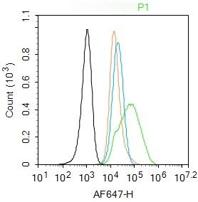 , ,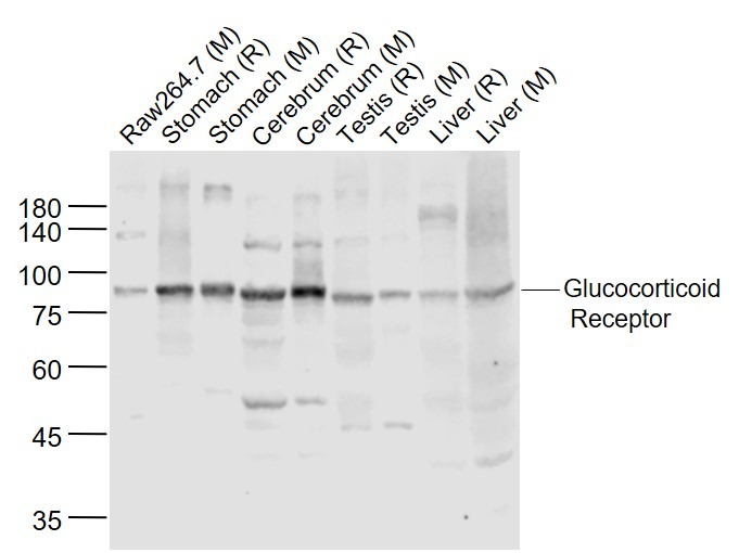 , ,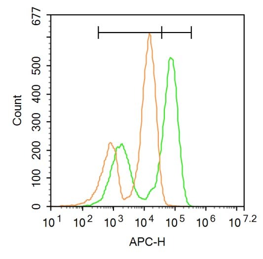 , ,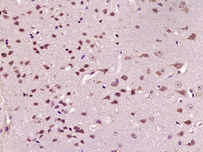 |
| Application | |
| Recommended Dose | WB: 1:500-2000; IHC-P: 1:100-500; IHC-Fr: 1:100-500; IF: 1:100-500; FCM: 1ug/Test |
| Antibody Type | Polyclonal |
| Host Species | Rabbit |
| Subcellular Localization | Cytoplasm. Nucleus. Note=Cytoplasmic in the absence of ligand, nuclear after ligand-binding. Isoform Beta: Nucleus. Note=Localized largely in the nucleus. |
| Tissue Specificity | Widely expressed. In the heart, detected in left and right atria, left and right ventricles, aorta, apex, intraventricular septum, and atrioventricular node as well as whole adult and fetal heart. |
| Construction | Polyclonal Antibody |
| Purification | Protein A purified |
| Appearance | Liquid |
| Formulation | 0.01M TBS (pH7.4) with 1% BSA, 0.02% Proclin300 and 50% Glycerol. |
| Concentration | 1 mg/mL |
| Research Background | Steroid receptors are ligand-dependent, intracellular proteins that stimulate transcription of specific genes by binding to specific DNA sequences following activation by the appropriate hormone. Glucocorticoids are a family of steroids necessary for the regulation of energy metabolism and the immune and inflammatory responses. These compounds exert their effect through their interaction with the glucocoticoid receptor (GR) and that complex's subsequent association with DNA. All normal mammalian tissues examined to date have been shown to contain glucocorticoid receptor. |
| Immunogen | KLH conjugated synthetic peptide: human Glucocorticoid Receptor beta |
| Antigen Species | Human |
| Gene Name | NR3C1 |
| Gene ID | |
| Protein Name | Glucocorticoid receptor |
| Uniprot ID | |
| Biology Area | Nuclear hormone receptors,Corticoid,Corticoid |
| Function | Receptor for glucocorticoids (GC). Has a dual mode of action: as a transcription factor that binds to glucocorticoid response elements (GRE) and as a modulator of other transcription factors. Affects inflammatory responses, cellular proliferation and differentiation in target tissues. Could act as a coactivator for STAT5-dependent transcription upon growth hormone (GH) stimulation and could reveal an essential role of hepatic GR in the control of body growth. Involved in chromatin remodeling. Plays a significant role in transactivation. Involved in nuclear translocation. |
| Molecular Weight | Theoretical: 86 kDa. Actual: 87 kDa. |
| Stability & Storage | Store at -20°C or -80°C for 12 months. Avoid repeated freeze-thaw cycles. |
| Transport | Shipping with blue ice. |
| Size | Quantity | Unit Price | Amount | Operation |
|---|
 Hello! How can I help you today?
 Copyright © 2015-2026 TargetMol Chemicals Inc. All Rights Reserved.
