
Shopping Cart
Remove All Your shopping cart is currently empty


Synonyms: RG7321, GDC-0941
| Pack Size | Price | USA Stock | Global Stock | Quantity |
|---|---|---|---|---|
| 5 mg | $38 | In Stock | In Stock | |
| 10 mg | $50 | In Stock | In Stock | |
| 50 mg | $64 | In Stock | In Stock | |
| 100 mg | $100 | In Stock | In Stock | |
| 200 mg | $180 | In Stock | In Stock | |
| 1 mL x 10 mM (in DMSO) | $50 | In Stock | In Stock |
| Description | Pictilisib (GDC-0941) (GDC-0941) is a potent pan inhibitor of class I catalytic subunits of PI3K (IC50s: 3/33/3/75 nM for p110α/β/δ/γ). |
| Targets & IC50 | p110α:3 nM (cell free), p110δ:3 nM (cell free), p110β:33 nM (cell free), p110α-E545K:3 nM, p110γ:75 nM, DNA-PK:1.23 μM, mTOR:0.58 μM (Ki), p110α-H1047R:3 nM |
| In vitro | Pictilisib is a potent inhibitor of cell proliferation in these cell lines with submicromolar IC50s. Potent inhibition of Akt (Ser473) phosphorylation was observed in U87MG, PC3, and MDA-MB-361 cells with IC50s of 46, 37, and 28 nM, respectively [1]. In comparison to single-agent treatments, the combination of Pictilisib and docetaxel reduced tumor cell viability by 80% or greater in the breast cancer cell lines tested in vitro. A Bliss sum of 0 was determined in the MDA-MB-453 cell line indicating an additive combination effect whereas Bliss sums > 0 were calculated in the other tumor cell lines indicating a synergistic effect [2]. Treatment with 250 nM Pictilisib for 2 hr resulted in 40%–85% inhibition of pAKT in all cell lines tested. Inhibition of the PI3K/AKT pathway by Pictilisib was reflected as a dose-dependent reduction in cell proliferation/viability. Pictilisib inhibited the growth of both trastuzumab-sensitive and -insensitive cells. The IC50 values for Pictilisib ranged between 150 and 950 nM and did not correlate with trastuzumab sensitivity [3]. |
| In vivo | Treatment of animals bearing MCF7-neo/HER2 breast cancer xenografts with 7.5 mg/kg docetaxel or 150 mg/kg Pictilisib led to tumor growth delay and tumor stasis, respectively. The combination of 100 mg/kg Pictilisib and docetaxel resulted in tumor stasis during the treatment period that was sustained after dosing ended [2]. AZD8055 (20mg/kg) or Pictilisib (75mg/kg) administration induced a transient increase in blood glucose levels. Treatment with either AZD8055 or Pictilisib led to a marked inhibition of Akt activity as well as phosphorylation of Thr308 and Ser473. Phosphorylation of the Akt substrates PRAS40 and Foxo-1/3a were also inhibited by AZD8055 or GDC-941 [4]. |
| Synonyms | RG7321, GDC-0941 |
| Kinase Assay | Recombinant human PI3Kα, PI3Kβ, and PI3Kδ are coexpressed in a Sf9 baculovirus system with the p85α regulatory subunit and purified as GST-fusion proteins using affinity chromatography on glutathione-sepharose. Recombinant human PI3Kγ is expressed as monomeric GST-fusions and purified similarly. GDC-0941 is dissolved in DMSO and added to 20 mM Tris-HCl (pH 7.5) containing 200 μg yttrium silicate (Ysi) polylysine SPA beads, 4 mM MgCl2, 1 mM dithiothreitol (DTT), 1 μM ATP, 0.125 μCi [γ-33P]-ATP, and 4% (v/v) DMSO in a total volume of 50 μL. The recombinant GST-fusion of PI3Kα (5 ng), PI3Kβ (5 ng), PI3Kδ (5 ng), or PI3Kγ (5 ng) is added to the assay mixture to initiate the kinase reaction. After incubation for 1 hour at room temperature, the kinase reaction is terminated with 150 μL PBS. The mixture is then centrifuged for 2 minutes at 2000 rpm and read using a Wallac Microbeta counter. The reported IC50 values are calculated using a sigmoidal, dose-response curve fit in MDL Assay Explorer [1]. |
| Cell Research | All drug treatments were tested in quadruplicate during a 4-day incubation period, and the relative number of viable cells was estimated using CellTiter-Glo. Total luminescence was measured on a Wallac Multilabel Reader. Cells were treated simultaneously with docetaxel (dose range = 0.0003–0.020 μmol/L) or GDC-0941 (dose range = 0.083–5 μmol/L) in an 8 × 10 matrix of concentrations chosen to encompass clinically relevant doses (24). The concentration of drug resulting in EC50 was determined using Prism software. Combination synergy of GDC-0941 and docetaxel was determined by Bliss independence analyses. A Bliss expectation for a combined response (C) was calculated by the equation: C = (A + B) ? (A × B) where A and B are the fractional growth inhibitions of drug A and B at a given dose. The difference between the Bliss expectation and the observed growth inhibition of the combination of drugs A and B at the same dose is the 'Delta.Bliss.' Delta.Bliss scores were summed across the dose matrix to generate a Bliss sum. Bliss sum = 0 indicates that the combination treatment is additive (as expected for independent pathway effects); Bliss sum > 0 indicates activity greater than additive (synergy); and Bliss sum < 0 indicates the combination is less than additive (antagonism). Statistical analysis comparing the Bliss sums for each cell line was conducted by the Student t-test [2]. |
| Animal Research | Female nu/nu mice were inoculated subcutaneously with MCF7-neo/HER2 or MX-1 breast cancer cells. When tumors reached a mean volume of 200 to 250 mm3, animals were size-matched and distributed into groups consisting of 10 animals per group. Docetaxel formulated in 3% EtOH, 97% saline was administered intravenously once weekly. GDC-0941, formulated in MCT (0.5% methylcellulose, 0.2% Tween-80) was dosed orally and daily. MAXF1162 is a HER2+/ER+/PR+ patient-derived breast cancer tumor xenograft model established by directly implanting tumors subcutaneously from patient to NMRI nu/nu mice. Tumor volume was calculated as follows: tumor size (mm3) = (longer measurement × shorter measurement2) × 0.5. Tumor sizes were recorded twice weekly over the course of a study. Following data analysis, P values were determined using the Dunnett t test. For pharmacodynamic studies, tumor samples (n = 4) were immediately frozen or fixed in 10% neutral-buffered formalin. Tumors were dissociated in cell extraction buffer, and lysates were analyzed by Western blotting as described above. Immunohistochemistry was conducted using 5-μm paraffin sections of formalin-fixed tissue on a Ventana Benchmark XT instrument by deparaffinization, treatment with antigen retrieval buffer, and incubation with anti-cleaved caspase-3 primary antibody at 37°C. Bound antibody was detected using DABMap technology, and sections were counterstained with hematoxylin [2]. |
| Molecular Weight | 513.64 |
| Formula | C23H27N7O3S2 |
| Cas No. | 957054-30-7 |
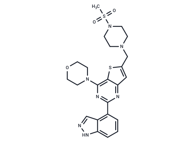
| Smiles | CS(=O)(=O)N1CCN(Cc2cc3nc(nc(N4CCOCC4)c3s2)-c2cccc3[nH]ncc23)CC1 |
| Relative Density. | 1.53 g/cm3 (Predicted) |
| Storage | Store at low temperature Powder: -20°C for 3 years | In solvent: -80°C for 1 year Shipping with blue ice/Shipping at ambient temperature. | |||||||||||||||||||||||||||||||||||
| Solubility Information | DMSO: 110.95 mg/mL (216.01 mM), Sonication is recommended. Ethanol: < 1 mg/mL (insoluble or slightly soluble) H2O: < 1 mg/mL (insoluble or slightly soluble) | |||||||||||||||||||||||||||||||||||
| In Vivo Formulation | 10% DMSO+40% PEG300+5% Tween 80+45% Saline: 4 mg/mL (7.79 mM), Sonication is recommended. Please add the solvents sequentially, clarifying the solution as much as possible before adding the next one. Dissolve by heating and/or sonication if necessary. Working solution is recommended to be prepared and used immediately. The formulation provided above is for reference purposes only. In vivo formulations may vary and should be modified based on specific experimental conditions. | |||||||||||||||||||||||||||||||||||
Solution Preparation Table | ||||||||||||||||||||||||||||||||||||
DMSO
Note : The dilution table applies only to solid products. For liquid products, please calculate the stock solution based on the stated concentration and/or density. | ||||||||||||||||||||||||||||||||||||
 For example, if the intended dosage is 10 mg/kg for animals weighing 20 g , with a dosing volume of 100 μL per animal, and a total of 10 animals are to be administered, using a formulation of
  10% DMSO+ 40% PEG300+ 5% Tween 80+ 45% Saline/PBS/ddH2O , the resulting working solution concentration would be 2 mg/mL.
Dissolve 2 mg of the compound in 100 μL DMSO to obtain a stock solution at a concentration of 20 mg/mL . If the required concentration exceeds the compound's known solubility, please contact us for technical support before proceeding.
1) Add 100 μL of the DMSO stock solution to 400 µL PEG300 and mix thoroughly until the solution becomes clear.
2) Add 50 µL Tween 80 and mix well until fully clarified.
3) Add 450 µL Saline,PBS or ddH2O and mix thoroughly until a homogeneous solution is obtained.
| Size | Quantity | Unit Price | Amount | Operation |
|---|
 Hello! How can I help you today?
 Copyright © 2015-2026 TargetMol Chemicals Inc. All Rights Reserved.
