
Shopping Cart
Remove All Your shopping cart is currently empty


Synonyms: OSI027, ASP4786

| Pack Size | Price | USA Stock | Global Stock | Quantity |
|---|---|---|---|---|
| 1 mg | $35 | In Stock | In Stock | |
| 2 mg | $48 | In Stock | In Stock | |
| 5 mg | $78 | In Stock | In Stock | |
| 10 mg | $141 | In Stock | In Stock | |
| 25 mg | $261 | In Stock | In Stock | |
| 50 mg | $409 | In Stock | In Stock | |
| 100 mg | $596 | In Stock | In Stock | |
| 1 mL x 10 mM (in DMSO) | $81 | In Stock | In Stock |
| Description | OSI-027 (ASP4786) is a selective and potent dual inhibitor of mTORC1 and mTORC2 with IC50 of 22 nM and 65 nM, and more than 100-fold selectivity observed for mTOR than PI3Kα, PI3Kβ, PI3Kγ or DNA-PK. Phase 1. |
| Targets & IC50 | mTOR:4 nM, PI3Kγ:0.42 μM, mTORC2:65 nM, DNA-PK:1 μM, mTORC1:22 nM, PI3Kα:1.3 μM |
| In vitro | OSI-027 shows the selective and ATP competitive inhibition activities against mTORC1 and mTORC2 with IC50 of 22 nM and 65 nM, respectively. In addition, OSI-027 inhibits mTOR signaling of phospho-4E-BP1 with an IC50 of 1 μM in cell-based assays. [1] OSI-027 exhibits anti-proliferative activities against several acute leukemia cell lines of myeloid/megakaryocytic origin in a dose-dependent manner, including U937, KG-1, KBM-3B, ML-1, HL-60, and MEG-01 cells. [2] A recent study shows that inhibition of mTORC1/2 by OSI-027 effectively suppresses phosphorylation of Akt (S473) and cell proliferation in breast cancer cells. [3] |
| In vivo | In GEO colorectal xenograft, OSI-027 (65 mg/kg) inhibits both mTORC1 and mTORC2 effectors, including 4E-BP1, Akt, and S6 phosphorylation. Furthermore, mTORC1 and mTORC2 inhibition together by OSI-027 potently inhibits tumor growth more than mTORC1 inhibition by rapamycin. [1] |
| Synonyms | OSI027, ASP4786 |
| Kinase Assay | Biochemical assays: mTORC1 and mTORC2 inhibition is assayed using native enzyme complex immunoprecipitated from HeLa lysates at 1 mM ATP. To prepare whole cell lysates from HeLa cells, 25 g cell pellet is lysed in 60 mL of ice-cold buffer A [40 mM HEPES (pH 7.5), 120 mM NaCl, 1 mM EDTA, 10 mM sodium pyrophosphate, 10 mM glycerophosphate, 50 mM NaF, 0.5 mM orthovanadate, and EDTA-free protease inhibitors containing 0.3% CHAPS] for 30 minutes on a magnetic stirrer in a cold room. After clearing of the lysates by centrifugation at 13,000 g for 10 minutes, Protein G-coated 384-well plates are incubated with 0.25 μg of mTOR antibody in 15 μL of buffer A for 1 hour at 4 °C. To each well, 40 μg of HeLa cell lysate in 15 μL of buffer A is added and incubated overnight at 4 °C to immunoprecipitate mTOR complexes. Plates are washed 3 times with buffer A and twice with immunoprecipitation wash buffer [Buffer B: 50 mM HEPES (pH 7.5) and 150 mM NaCl]. OSI-027 is added at 10 μM concentration to each well and DMSO is added to the control wells. The reaction is started by adding 150 ng of His-tagged 4E-BP1 as a substrate in the presence or absence of 100 μM ATP to each well in 25 μL of freshly prepared kinase buffer [Buffer C: 20 mM HEPES (pH 7.5), 10 mM MgCl2, 4 mM MnCl2, 10 mM β-mercaptoethanol, and 200 μM vanadate] and incubated at room temperature (RT) for 30 minutes. The reaction is stopped by transferring 25 μL of reaction mixture from each well to corresponding wells of fresh Ni-chelate-coated plates and incubated overnight at 4 °C followed by 2 hours at 37 °C. To detect phosphorylation of 4E-BP1, the plates are washed once with TBST (Tris-buffered saline containing 0.1% Tween-20) containing 5% skim milk powder. To each well, 25 μL of 1:1,000 diluted phospho-4E-BP1 antibodies in TBST containing 5% skim milk are added and incubated for 1 hour at RT.The plates are washed once with TBST and then 25 μL of anti-rabbit HRP (diluted 1:10,000) in TBST containing 5% skim milk is added. The plates are incubated for 1 hour at RT and washed 5 times with TBST. For detection of phospho-4E-BP1, 25 μL of chemiluminescent reagents A+B is added and chemiluminescence is measured using an Analyst plate reader. |
| Cell Research | Inhibition of proliferation is measured using the Cell Titer Glo Assay , as noted in figure legends. To generate dose–response curves, cell lines are seeded at a density of 5,000 cells per well in a 96-well plate. After 24 hours of plating, cells are dosed with varying concentrations of either OSI-027 or rapamycin. The signal for Cell Titer Glo Assay is determined 72 hours after dosing and normalized to that of vehicle-treated controls. Inhibition of proliferation, relative to vehicle-treated controls, is expressed as a fraction of 1 and graphed using PRISM software. (Only for Reference) |
| Molecular Weight | 406.44 |
| Formula | C21H22N6O3 |
| Cas No. | 936890-98-1 |
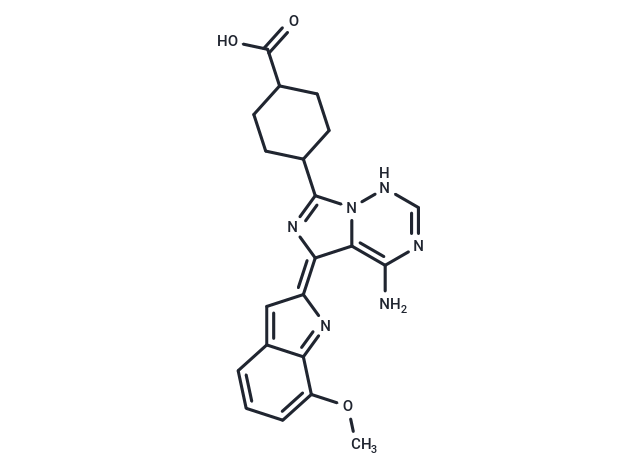
| Smiles | COc1cccc2=C\C(N=c12)=c1/nc(C2CCC(CC2)C(O)=O)n2NC=NC(N)=c12 |
| Relative Density. | 1.59 g/cm3 |
| Storage | Powder: -20°C for 3 years | In solvent: -80°C for 1 year Shipping with blue ice/Shipping at ambient temperature. | |||||||||||||||||||||||||
| Solubility Information | DMSO: 15 mg/mL (36.91 mM), Sonication is recommended. H2O: < 1 mg/mL (insoluble or slightly soluble) Ethanol: < 1 mg/mL (insoluble or slightly soluble) | |||||||||||||||||||||||||
| In Vivo Formulation | 10% DMSO+40% PEG300+5% Tween 80+45% Saline: 2 mg/mL (4.92 mM), Sonication is recommended. Please add the solvents sequentially, clarifying the solution as much as possible before adding the next one. Dissolve by heating and/or sonication if necessary. Working solution is recommended to be prepared and used immediately. The formulation provided above is for reference purposes only. In vivo formulations may vary and should be modified based on specific experimental conditions. | |||||||||||||||||||||||||
Solution Preparation Table | ||||||||||||||||||||||||||
DMSO
Note : The dilution table applies only to solid products. For liquid products, please calculate the stock solution based on the stated concentration and/or density. | ||||||||||||||||||||||||||
 For example, if the intended dosage is 10 mg/kg for animals weighing 20 g , with a dosing volume of 100 μL per animal, and a total of 10 animals are to be administered, using a formulation of
  10% DMSO+ 40% PEG300+ 5% Tween 80+ 45% Saline/PBS/ddH2O , the resulting working solution concentration would be 2 mg/mL.
Dissolve 2 mg of the compound in 100 μL DMSO to obtain a stock solution at a concentration of 20 mg/mL . If the required concentration exceeds the compound's known solubility, please contact us for technical support before proceeding.
1) Add 100 μL of the DMSO stock solution to 400 µL PEG300 and mix thoroughly until the solution becomes clear.
2) Add 50 µL Tween 80 and mix well until fully clarified.
3) Add 450 µL Saline,PBS or ddH2O and mix thoroughly until a homogeneous solution is obtained.
| Size | Quantity | Unit Price | Amount | Operation |
|---|
 Hello! How can I help you today?
 Copyright © 2015-2026 TargetMol Chemicals Inc. All Rights Reserved.
