
Shopping Cart
Remove All Your shopping cart is currently empty


Synonyms: KU0059436, AZD2281
| Pack Size | Price | USA Stock | Global Stock | Quantity |
|---|---|---|---|---|
| 5 mg | $31 | In Stock | In Stock | |
| 10 mg | $48 | In Stock | In Stock | |
| 50 mg | $68 | In Stock | In Stock | |
| 100 mg | $91 | In Stock | In Stock | |
| 200 mg | $120 | In Stock | In Stock | |
| 500 mg | $179 | In Stock | In Stock | |
| 1 g | $247 | In Stock | In Stock |
| Description | Olaparib (KU0059436) is a small molecule inhibitor of PARP1/PARP2 (IC50=5/1 nM), with weak inhibitory activity against PARP tankyrase-1 (IC50=1.5 μM), and is selective and orally active. Olaparib exhibits autophagy and mitochondrial autophagy activation activity. |
| Targets & IC50 | PARP1:5 nM (cell free), tankyrase 1:1.5 μM, PARP2:1 nM (cell free) |
| In vitro | METHODS: Human cervical cancer cells SiHa and ME180 were treated with Olaparib (5-10 µM) and cisplatin (1-30 µM) for 72 h. Cell growth inhibition was detected by MTT. RESULTS: Olaparib and cisplatin co-treatment showed significant cell growth inhibition compared to cells treated with a single drug. [1] METHODS: Human endometrial cancer cells HEC-6 and HEC-6-PTEN were treated with Olaparib (10 μM) for 72 h. The cell cycle was analyzed by Flow Cytometry. RESULTS: Olaparib induced a significant increase in the sub-G1 population of HEC-6 and HEC-6-PTEN cells. [2] METHODS: Chicken lymphoma cells DT40 were treated with Olaparib (0.01-10 μM) for 30 min, and the expression levels of target proteins were detected by Western Blot. RESULTS: Olaparib dose-dependently inhibited the expression level of PARylation and the activation of PARP. [3] |
| In vivo | METHODS: To detect anti-tumor activity in vivo, Olaparib (10 mg/kg) and TMZ (50 mg/kg) were orally administered to mice bearing human colorectal cancer tumor SW620 once daily for five days. RESULTS: Significant suppression of tumor volume was observed in the TMZ plus Olaparib combination treatment group compared to the TMZ group alone. [4] METHODS: To investigate the therapeutic effects of Olaparib on asthma, Olaparib (1-10 mg/kg) was administered intraperitoneally once daily for three days to an OVA-based asthmatic C57BL/6 mouse model. RESULTS: Olaparib significantly reduced airway eosinophilia, mucus production, and hyperresponsiveness. The protective effects of Olaparib were associated with inhibition of the Th2 cytokines eotaxin, IL-4, IL-5, IL-6, IL-13, and M-CSF, as well as ovalbumin-specific IgE, and an increase in the Th1 cytokine IFN-γ. Olaparib is a potential candidate for clinical trials in human asthma. [5] |
| Synonyms | KU0059436, AZD2281 |
| Kinase Assay | This assay determined the ability of test compounds to inhibit PARP-1 enzyme activity. The method that was used was as reported. We measured PARP-2 activity inhibition by using a variation of the PARP-1 assay in which PARP-2 protein (recombinant) was bound down by a PARP-2 specific antibody in a 96-well white-walled plate. PARP-2 activity was measured following 3H-NAD+ DNA additions. After washing, scintillant was added to measure 3 H-incorporated ribosylations. For tankyrase-1, an AlphaScreen assay was developed in which HIS-tagged recombinant TANK-1 protein was incubated with biotinylated NAD+ in a 384-well ProxiPlate assay. Alpha beads were added to bind the HIS and biotin tags to create a proximity signal, whereas the inhibition of TANK-1 activity was directly proportional to the loss of this signal. All experiments were repeated at least three times [1]. |
| Cell Research | HSC-2, Ca9-22, and SAS oral carcinoma cells were seeded in 24-well plates at a density of 2 × 10⁴ cells/well. After overnight incubation, the culture medium was replaced with fresh medium containing various concentrations of PARP inhibitor AZD228 or cisplatin. After 24 h of treatment, the number of viable cells was assessed using an MTT assay as reported previously. Briefly, one-tenth of the fluid volume of 5 mg/mL MTT in RPMI-1640 medium was added to each well, followed by incubation for 4 h at 37 °C. After incubation, the medium was carefully removed and an adequate volume of 0.1 N HCl in isopropanol was added to each well and the resultant formazan crystals was dissolved. Absorbance was determined at 570 nm by microplate reader in 96-well assay plates. All experiments were performed in triplicate [2]. |
| Animal Research | Once the tumor diameter had reached 7 mm, the mice were randomly assigned to the following groups: (a) control (200 μL saline); (b) cisplatin (2 mg/kg per body weight, dissolved in 200 μL sterilized water); (c) AZD2281 (25 mg/kg per body weight, dissolved in 200 μL sterilized water); or (d) combination (both cisplatin and AZD2281). The chemicals were administered intraperitoneally every three days, five times. Although AZD2281 is administered orally in the clinic, intraperitoneal injection was recommended by the manufacturer because of easier manipulation and the ethical constraints associated with oral gavage administration to mice. Tumor size and body weight were measured at the time of administration. The tumor volume was calculated using following equation. Tumor volume = verticality × width × height × 0.5236. Three days after the last administration, all surviving mice were sacrificed [2]. |
| Molecular Weight | 434.46 |
| Formula | C24H23FN4O3 |
| Cas No. | 763113-22-0 |
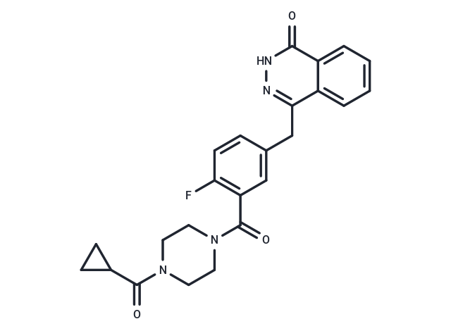
| Smiles | C(C=1C=2C(C(=O)NN1)=CC=CC2)C3=CC(C(=O)N4CCN(C(=O)C5CC5)CC4)=C(F)C=C3 |
| Relative Density. | 1.43 |
| Storage | Powder: -20°C for 3 years | In solvent: -80°C for 1 year Shipping with blue ice/Shipping at ambient temperature. | |||||||||||||||||||||||||||||||||||
| Solubility Information | H2O: < 1 mg/mL (insoluble) DMSO: 82.5 mg/mL (189.89 mM), Sonication is recommended. Ethanol: < 1 mg/mL (insoluble or slightly soluble) | |||||||||||||||||||||||||||||||||||
| In Vivo Formulation | 10% DMSO+40% PEG300+5% Tween 80+45% Saline: 8 mg/mL (18.41 mM), Solution. Please add the solvents sequentially, clarifying the solution as much as possible before adding the next one. Dissolve by heating and/or sonication if necessary. Working solution is recommended to be prepared and used immediately. The formulation provided above is for reference purposes only. In vivo formulations may vary and should be modified based on specific experimental conditions. | |||||||||||||||||||||||||||||||||||
Solution Preparation Table | ||||||||||||||||||||||||||||||||||||
DMSO
Note : The dilution table applies only to solid products. For liquid products, please calculate the stock solution based on the stated concentration and/or density. | ||||||||||||||||||||||||||||||||||||
 For example, if the intended dosage is 10 mg/kg for animals weighing 20 g , with a dosing volume of 100 μL per animal, and a total of 10 animals are to be administered, using a formulation of
  10% DMSO+ 40% PEG300+ 5% Tween 80+ 45% Saline/PBS/ddH2O , the resulting working solution concentration would be 2 mg/mL.
Dissolve 2 mg of the compound in 100 μL DMSO to obtain a stock solution at a concentration of 20 mg/mL . If the required concentration exceeds the compound's known solubility, please contact us for technical support before proceeding.
1) Add 100 μL of the DMSO stock solution to 400 µL PEG300 and mix thoroughly until the solution becomes clear.
2) Add 50 µL Tween 80 and mix well until fully clarified.
3) Add 450 µL Saline,PBS or ddH2O and mix thoroughly until a homogeneous solution is obtained.
| Size | Quantity | Unit Price | Amount | Operation |
|---|
 Hello! How can I help you today?
 Copyright © 2015-2026 TargetMol Chemicals Inc. All Rights Reserved.
