
Shopping Cart
Remove All Your shopping cart is currently empty


Synonyms: AG-013736
| Pack Size | Price | USA Stock | Global Stock | Quantity |
|---|---|---|---|---|
| 25 mg | $33 | In Stock | In Stock | |
| 50 mg | $45 | In Stock | In Stock | |
| 100 mg | $61 | In Stock | In Stock | |
| 200 mg | $77 | In Stock | In Stock | |
| 500 mg | $150 | In Stock | In Stock | |
| 1 g | $218 | In Stock | In Stock | |
| 1 mL x 10 mM (in DMSO) | $48 | In Stock | In Stock |
| Description | Axitinib (AG-013736) is a multi-targeted tyrosine kinase inhibitor that inhibits VEGFR1, VEGFR2, VEGFR3, and PDGFRβ (IC50=4/20/0.4/2 nM). Axitinib has antitumor activity and is used in the treatment of renal cell carcinoma. |
| Targets & IC50 | PDGFRβ:1.6 nM, VEGFR2/KDR:0.2 nM, VEGFR1/FLT1:0.1 nM, VEGFR2/Flk1:0.18 nM, c-Kit:1.7 nM, VEGFR3:0.1-0.3 nM |
| In vitro | METHODS: Thirteen neuroblastoma cells were treated with Tepotinib for 72 h and cell viability was measured by MTT assay. RESULTS: All cells showed reduced cell viability with IC50 values ranging from 2.4-8.5 µM.[1] METHODS: EBC-1 cells were treated with Tepotinib (0-30 µmol/L) for 3 days, and the expression levels of target proteins were detected by Western Blot. RESULTS: Tepotinib treatment induced a significant reduction in c-Met constitutive phosphorylation in EBC-1 cells with an IC50 of 9 nmol/L. Tepotinib also effectively blocked the phosphorylation of the major downstream effectors of the c-Met enzyme in EBC-1, MKN-45, and Hs746T cells in the 1-10 nmol/L range. [2] |
| In vivo | METHODS: To assay antitumor activity in vivo, Tepotinib (5-15 mg/kg) was injected once daily for 14 days into CD-1 mice bearing EBC-1 xenografts. RESULTS: Administration of 5 or 15 mg/kg of Tepotinib daily to mice bearing EBC-1 tumors effectively inhibited or completely regressed the tumors, respectively. [2] |
| Synonyms | AG-013736 |
| Kinase Assay | Porcine aorta endothelial (PAE) cells overexpressing full-length VEGFR-2, PDGFR-β, KIT, and NIH-3T3 overexpressing murine VEGFR-2 (Flk-1) or PDGFR-α were generated as described previously. The ELISA capture plates were prepared by coating 96-well ReactiBind plates with 100 μL/well of 2.5 μg/mL anti-VEGFR-2 antibody, 0.75 μg/mL anti-PDGFR-β antibody, 0.25 μg/mL anti-PDGFR-α antibody, 0.5 μg/mL anti-KIT antibody, or 1.20 μg/mL anti-Flk-1 antibody. Measurement of RTK phosphorylation by ELISA was done as described previously [1]. |
| Cell Research | Endothelial or tumor cells were starved for 18 h in the presence of either 1% FBS (HUVEC) or 0.1% FBS (tumor cells). Axitinib was added and cells were incubated for 45 min at 37°C in the presence of 1 mmol/L Na3VO4. The appropriate growth factor was added to the cells, and after 5 min, cells were rinsed with cold PBS and lysed in the lysis buffer and a protease inhibitor cocktail. The lysates were incubated with immunoprecipitation antibodies for the intended proteins overnight at 4°C. Antibody complexes were conjugated to protein A beads and supernatants were separated by SDS-PAGE. The Super Signal West Dura kit was used to detect the chemiluminescent signal [1]. |
| Animal Research | AG-013736, a receptor kinase inhibitor of VEGFRs and, at higher doses, PDGFRs (IC50 = 0.1 nmol/L for VEGFR-1, 0.2 nmol/L for VEGFR-2, 0.1–0.3 nmol/L for VEGFR-3, and 1.6 nmol/L for PDGFRβ; ref. 18), was provided by Pfizer Global Research and given once daily by gavage in a volume of 0.13 mL. Control animals received 0.5% carboxymethylcellulose drug carrier. Irradiations were done on nonanesthetized mice using a 137Cs source operating at 2.4 Gy/min. Mice were confined to plastic jigs with tumor-bearing legs extended through an opening in the side, allowing local irradiations. Fractionated doses were given in five daily 2 Gy fractions per week (omitting weekends). For combination treatments, radiotherapy was delivered first, and AG-013736 was given within ~4 h. Mice were sacrificed, and tumors were excised and then quick frozen (using liquid nitrogen) following 1, 2, or 3 weeks of treatment [3]. |
| Molecular Weight | 386.47 |
| Formula | C22H18N4OS |
| Cas No. | 319460-85-0 |
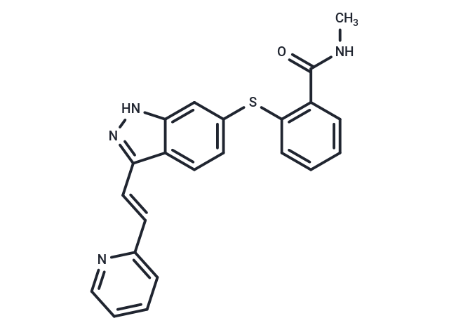
| Smiles | C(=C/C1=CC=CC=N1)\C=2C=3C(=CC(SC4=C(C(NC)=O)C=CC=C4)=CC3)NN2 |
| Relative Density. | 1.35 g/cm3 |
| Storage | Store at low temperature,Keep away from moisture,Keep away from direct sunlight Powder: -20°C for 3 years | In solvent: -80°C for 1 year Shipping with blue ice/Shipping at ambient temperature. | ||||||||||||||||||||||||||||||
| Solubility Information | DMSO: 23.2 mg/mL (60.03 mM), Sonication is recommended. | ||||||||||||||||||||||||||||||
| In Vivo Formulation | 10% DMSO+40% PEG300+5% Tween 80+45% Saline: 2 mg/mL (5.18 mM), Sonication is recommended. Please add the solvents sequentially, clarifying the solution as much as possible before adding the next one. Dissolve by heating and/or sonication if necessary. Working solution is recommended to be prepared and used immediately. The formulation provided above is for reference purposes only. In vivo formulations may vary and should be modified based on specific experimental conditions. | ||||||||||||||||||||||||||||||
Solution Preparation Table | |||||||||||||||||||||||||||||||
DMSO
Note : The dilution table applies only to solid products. For liquid products, please calculate the stock solution based on the stated concentration and/or density. | |||||||||||||||||||||||||||||||
 For example, if the intended dosage is 10 mg/kg for animals weighing 20 g , with a dosing volume of 100 μL per animal, and a total of 10 animals are to be administered, using a formulation of
  10% DMSO+ 40% PEG300+ 5% Tween 80+ 45% Saline/PBS/ddH2O , the resulting working solution concentration would be 2 mg/mL.
Dissolve 2 mg of the compound in 100 μL DMSO to obtain a stock solution at a concentration of 20 mg/mL . If the required concentration exceeds the compound's known solubility, please contact us for technical support before proceeding.
1) Add 100 μL of the DMSO stock solution to 400 µL PEG300 and mix thoroughly until the solution becomes clear.
2) Add 50 µL Tween 80 and mix well until fully clarified.
3) Add 450 µL Saline,PBS or ddH2O and mix thoroughly until a homogeneous solution is obtained.
| Size | Quantity | Unit Price | Amount | Operation |
|---|
 Hello! How can I help you today?
 Copyright © 2015-2026 TargetMol Chemicals Inc. All Rights Reserved.
