
Shopping Cart
Remove All Your shopping cart is currently empty

Synonyms: PDGFR-β, PDGFRB, PDGFR-2, PDGFR1, PDGFR, PDGF R β/CD140b, PDGF R β, PDGF R beta, JTK12, CD140B
Anti-PDGF R beta/CD140b Polyclonal Antibody
| Pack Size | Price | USA Stock | Global Stock | Quantity |
|---|---|---|---|---|
| 50 µL | $222 | 7-10 days | 7-10 days | |
| 100 µL | $372 | 7-10 days | 7-10 days | |
| 200 µL | $527 | 7-10 days | 7-10 days |
| Description | Anti-PDGF R beta/CD140b Polyclonal Antibody is a Rabbit antibody targeting PDGF R beta/CD140b. Anti-PDGF R beta/CD140b Polyclonal Antibody can be used in IF,IHC-Fr,WB. |
| Synonyms | PDGFR-β, PDGFRB, PDGFR-2, PDGFR1, PDGFR, PDGF R β/CD140b, PDGF R β, PDGF R beta, JTK12, CD140B |
| Ig Type | IgG |
| Reactivity | Human,Rat (predicted:Mouse,Dog,Cow) |
| Verified Activity | 1. Sample: DU145 (Human) Cell Lysate at 30 μg Primary: Anti-PDGF Receptor beta (TMAB-01350) at 1/300 dilution Secondary: IRDye800CW Goat Anti-Rabbit IgG at 1/20000 dilution Predicted band size: 118 kDa Observed band size: 118 kDa 2. Sample: U251 (Human) Cell Lysate at 30 μg Primary: Anti-PDGF Receptor beta (TMAB-01350) at 1/300 dilution Secondary: IRDye800CW Goat Anti-Rabbit IgG at 1/20000 dilution Predicted band size: 118 kDa Observed band size: 118 kDa 3. Sample: NIH/3T3 (Rat) Cell Lysate at 30 μg Primary: Anti-PDGF Receptor beta (TMAB-01350) at 1/300 dilution Secondary: IRDye800CW Goat Anti-Rabbit IgG at 1/20000 dilution Predicted band size: 118 kDa Observed band size: 118 kDa 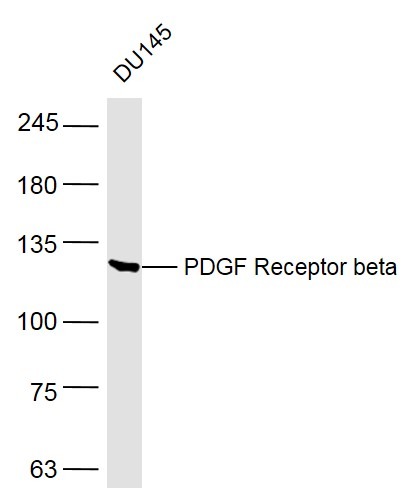 , ,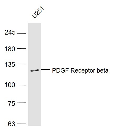 , ,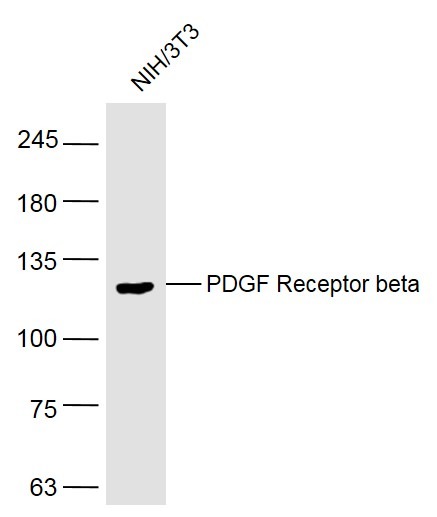 |
| Application | |
| Recommended Dose | WB: 1:500-2000; IHC-Fr: 1:100-500; IF: 1:100-500 |
| Antibody Type | Polyclonal |
| Host Species | Rabbit |
| Subcellular Localization | Cell membrane; Single-pass type I membrane protein. Cytoplasmic vesicle. Lysosome lumen. Note=After ligand binding, the autophosphorylated receptor is ubiquitinated and internalized, leading to its degradation |
| Construction | Polyclonal Antibody |
| Purification | Protein A purified |
| Appearance | Liquid |
| Formulation | 0.01M TBS (pH7.4) with 1% BSA, 0.02% Proclin300 and 50% Glycerol. |
| Concentration | 1 mg/mL |
| Research Background | This gene encodes a cell surface tyrosine kinase receptor for members of the platelet-derived growth factor family. These growth factors are mitogens for cells of mesenchymal origin. The identity of the growth factor bound to a receptor monomer determines whether the functional receptor is a homodimer or a heterodimer, composed of both platelet-derived growth factor receptor alpha and beta polypeptides. This gene is flanked on chromosome 5 by the genes for granulocyte-macrophage colony-stimulating factor and macrophage-colony stimulating factor receptor; all three genes may be implicated in the 5-q syndrome. A translocation between chromosomes 5 and 12, that fuses this gene to that of the translocation, ETV6, leukemia gene, results in chronic myeloproliferative disorder with eosinophilia. [provided by RefSeq]. |
| Immunogen | KLH conjugated synthetic peptide: human PDGF-R-B |
| Antigen Species | Human |
| Gene Name | PDGFRB |
| Gene ID | |
| Protein Name | Platelet-derived growth factor receptor beta |
| Uniprot ID | |
| Biology Area | PDGF,Growth factor receptors,Growth Factors,Inflammatory mediators,SARS Coronavirus,PDGF,Receptor Tyrosine Kinases |
| Function | Tyrosine-protein kinase that acts as cell-surface receptor for homodimeric PDGFB and PDGFD and for heterodimers formed by PDGFA and PDGFB, and plays an essential role in the regulation of embryonic development, cell proliferation, survival, differentiation, chemotaxis and migration. Plays an essential role in blood vessel development by promoting proliferation, migration and recruitment of pericytes and smooth muscle cells to endothelial cells. Plays a role in the migration of vascular smooth muscle cells and the formation of neointima at vascular injury sites. Required for normal development of the cardiovascular system. Required for normal recruitment of pericytes (mesangial cells) in the kidney glomerulus, and for normal formation of a branched network of capillaries in kidney glomeruli. Promotes rearrangement of the actin cytoskeleton and the formation of membrane ruffles. Binding of its cognate ligands - homodimeric PDGFB, heterodimers formed by PDGFA and PDGFB or homodimeric PDGFD -leads to the activation of several signaling cascades; the response depends on the nature of the bound ligand and is modulated by the formation of heterodimers between PDGFRA and PDGFRB. Phosphorylates PLCG1, PIK3R1, PTPN11, RASA1/GAP, CBL, SHC1 and NCK1. Activation of PLCG1 leads to the production of the cellular signaling molecules diacylglycerol and inositol 1,4,5-trisphosphate, mobilization of cytosolic Ca(2+) and the activation of protein kinase C. Phosphorylation of PIK3R1, the regulatory subunit of phosphatidylinositol 3-kinase, leads to the activation of the AKT1 signaling pathway. Phosphorylation of SHC1, or of the C-terminus of PTPN11, creates a binding site for GRB2, resulting in the activation of HRAS, RAF1 and down-stream MAP kinases, including MAPK1/ERK2 and/or MAPK3/ERK1. Promotes phosphorylation and activation of SRC family kinases. Promotes phosphorylation of PDCD6IP/ALIX and STAM. Receptor signaling is down-regulated by protein phosphatases that dephosphorylate the receptor and its down-stream effectors, and by rapid internalization of the activated receptor. |
| Molecular Weight | Theoretical: 118 kDa. Actual: 118 kDa. |
| Stability & Storage | Store at -20°C or -80°C for 12 months. Avoid repeated freeze-thaw cycles. |
| Transport | Shipping with blue ice. |
| Size | Quantity | Unit Price | Amount | Operation |
|---|
 Hello! How can I help you today?
 Copyright © 2015-2026 TargetMol Chemicals Inc. All Rights Reserved.
