
Shopping Cart
Remove All Your shopping cart is currently empty


Synonyms: Ammoniated ruthenium oxychloride
| Pack Size | Price | USA Stock | Global Stock | Quantity |
|---|---|---|---|---|
| 100 mg | $34 | In Stock | In Stock | |
| 200 mg | $48 | In Stock | In Stock |
| Description | Ruthenium Red (Ammoniated ruthenium oxychloride) is a polycationic dye widely used for electron microscopy (EM) of cells, tissues and vegetative bacteria. Ruthenium red strongly reacts with phospholipids and fatty acids and binds to acidic mucopolysaccharides. Ruthenium red is a L-type calcium current (ICa) blocker, blocks Ca2+ uptake and release, and voltage-sensitive Ca2+ channels |
| Synonyms | Ammoniated ruthenium oxychloride |
| Cell Research | 1. Solution preparation 1. Mother solution preparation: Dissolve Ruthenium Red in an appropriate solvent, DMSO or H2O, usually at a concentration of 1-10 mM. 2. Working solution preparation: Dilute Ruthenium Red into the required working solution using PBS/DMEM/H2O, usually at a concentration range of 1-10μM, which needs to be adjusted according to the experimental situation. 1. As a calcium current blocker 1. Cell treatment: Add Ruthenium Red working solution to the cell culture medium and incubate at different time points to observe the inhibitory effect of L-type calcium channels. 2. Calcium ion monitoring: Use calcium indicators (such as Fura-2, Fluo-4, etc.) combined with fluorescence microscopy or flow cytometry to monitor changes in intracellular calcium ion concentration. 2. For electron microscopy observation 1. Sample staining: Add Ruthenium Red working solution to cells or tissue samples, stain for a certain period of time, remove and wash with PBS. 2. Microscopic observation: Observe the sample using an electron microscope or fluorescence microscope to check the staining effect. III. Cell membrane and tissue staining: 1. Solution preparation: Dilute the Ruthenium Red stock solution in PBS or other suitable buffer, and the concentration can be adjusted to 10-50 μM. 2. Cell or tissue staining: Add the dye solution to the cell culture medium, incubate for 15-30 minutes, and then observe the labeled cell structure by fluorescence microscopy. The above information is based on published literature. Experimental procedures should be appropriately modified to meet specific research demands. |
| Molecular Weight | 786.34 |
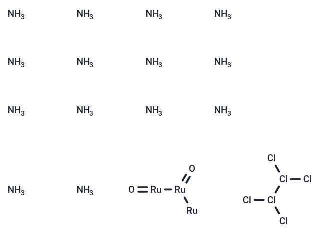
| Formula | Cl6H42N14O2Ru3 |
| Cas No. | 11103-72-3 |
| Smiles | N.N.N.N.N.N.N.N.N.N.N.N.N.N.[Ru][Ru](=O)[Ru]=O.Cl[Cl](Cl)[Cl](Cl)Cl |
| Relative Density. | 3.11 |
| Storage | Keep away from direct sunlight,Store under nitrogen Powder: -20°C for 3 years | In solvent: -80°C for 1 year Shipping with blue ice/Shipping at ambient temperature. | |||||||||||||||||||||||||
| Solubility Information | H2O: 20 mg/mL (25.43 mM), Sonication is recommended. DMSO: 20 mg/mL (25.43 mM), Sonication is recommended. | |||||||||||||||||||||||||
Solution Preparation Table | ||||||||||||||||||||||||||
H2O/DMSO
Note : The dilution table applies only to solid products. For liquid products, please calculate the stock solution based on the stated concentration and/or density. | ||||||||||||||||||||||||||
 For example, if the intended dosage is 10 mg/kg for animals weighing 20 g , with a dosing volume of 100 μL per animal, and a total of 10 animals are to be administered, using a formulation of
  10% DMSO+ 40% PEG300+ 5% Tween 80+ 45% Saline/PBS/ddH2O , the resulting working solution concentration would be 2 mg/mL.
Dissolve 2 mg of the compound in 100 μL DMSO to obtain a stock solution at a concentration of 20 mg/mL . If the required concentration exceeds the compound's known solubility, please contact us for technical support before proceeding.
1) Add 100 μL of the DMSO stock solution to 400 µL PEG300 and mix thoroughly until the solution becomes clear.
2) Add 50 µL Tween 80 and mix well until fully clarified.
3) Add 450 µL Saline,PBS or ddH2O and mix thoroughly until a homogeneous solution is obtained.
| Size | Quantity | Unit Price | Amount | Operation |
|---|
 Hello! How can I help you today?
 Copyright © 2015-2026 TargetMol Chemicals Inc. All Rights Reserved.
