
Shopping Cart
Remove All Your shopping cart is currently empty

Synonyms: EI 275, AGL 1872

| Pack Size | Price | USA Stock | Global Stock | Quantity |
|---|---|---|---|---|
| 1 mg | $34 | In Stock | In Stock | |
| 2 mg | $48 | In Stock | In Stock | |
| 5 mg | $81 | In Stock | In Stock | |
| 10 mg | $121 | In Stock | In Stock | |
| 25 mg | $212 | In Stock | In Stock | |
| 50 mg | $297 | In Stock | In Stock | |
| 100 mg | $468 | In Stock | In Stock | |
| 500 mg | $1,050 | - | In Stock | |
| 1 mL x 10 mM (in DMSO) | $86 | In Stock | In Stock |
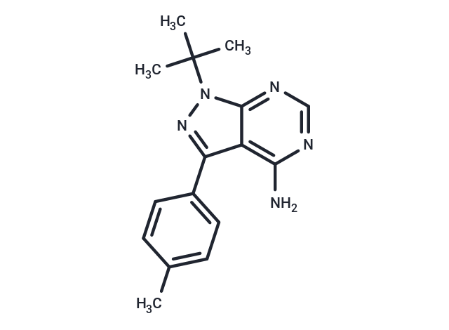
| Description | PP1 (AGL 1872), a specific and effective Src inhibitor, is with IC50 for Lck/Fyn is 5 nM/ 6 nM, respectively. |
| Targets & IC50 | Lck:5 nM, Fyn:6 nM |
| In vitro | PP1 is a nano-molar inhibitor of Lck and FynT, inhibits anti-CD3-induced protein-tyrosine kinase activity in T cells (IC50, 0.5 μM), demonstrates selectivity for Lck and FynT over ZAP-70, and preferentially inhibits T cell receptor-dependent anti-CD3-induced T cell proliferation (IC50, 0. 5 μM) over non-T cell receptor-dependent phorbol 12-myristate 13-acetate/interleu-kin-2 (IL-2)-induced T cell proliferation. PP1 (1 μM) selectively inhibits the induction of the IL-2 gene, but not the granulocyte-macrophage colony-stimulating factor or IL-2 receptor genes. PP1 also inhibits Src (IC50, 170 nM) and Hck (IC50, 20 nM). PP1 is 50–100-fold less active in the inhibition of A-431 epidermal growth factor receptor autophosphorylation (IC50, 0.25 μM). [1] PP1 also inhibits Kit and Bcr-Abl tyrosine kinases with IC50 of ~75 nM and 1 μM, respectively. PP1 completely abrogates the proliferation of M07e cells in response to SCF with IC50 of 0.5–1 μM. PP1 (1 μM) inhibits SCF-induced c-Kit autophosphorylation in intact cells and blocks the activation of mitogen-activated protein kinase and Akt. PP1 inhibits the activity of mutant constitutively active forms of c-Kit (D814V and D814Y) found in mast cell disorders, and triggers apoptosis in the rat basophilic leukemia cell line RBL-2H3 that expresses mutant c-Kit. PP1 reduces the constitutive activation of signal transducer and activators of transcription 5 and mitogen-activated protein kinase and triggeres apoptosis in FDCP1 cells expressing Bcr-Abl. [2] |
| Synonyms | EI 275, AGL 1872 |
| Kinase Assay | Protein A-Sepharose beads (prepared as a 50% (w/v) suspension) are added to the antibody/lysate mixture at 250 μL/mL and allowed to incubate for 30 min at 4°C. The beads are then washed twice in 1 mL of lysis buffer and twice in 1 mL of kinase buffer (25 mM HEPES, 3 mM MnCl2, 5 mM MgCl2, and 100 μM sodium orthovanadate) and resuspended to 50% (w/v) in kinase buffer. Twenty-five microliters of the bead suspension is added to each well of the enolase-coated 96-well high protein binding plate together with an appropriate concentration of compound and [γ-32P]ATP (25 μL/well of a 200 μCi/mL solution in kinase buffer). After incubation for 20 min at 20°C, 60 μL of boiling 2× solubilization buffer containing 10 mM ATP is added to the assay wells to terminate the reactions. Thirty microliters of the samples is removed from the wells, boiled for 5 min, and run on a 7.5% SDS-polyacrylamide gel. The gels are subsequently dried and exposed to Kodak X-AR film. For quantitation, films are scanned using a Molecular Dynamics laser scanner, and the optical density of the major substrate band, enolase p46, is determined. Concentrations of compound that causes 50% inhibition of enolase phosphorylation (IC50) are determined from a plot of the density versus concentration of compound. In companion experiments for measuring the activity of compounds against Lck, the assay plate is washed with two wash cycles on a Skatron harvester using 50 mM EDTA, 1 mM ATP. Scintillation fluid (100 μL) is then added to the wells, and P incorporation is measured using a Pharmacia Biotech micro-β-counter. Concentrations of compound that causes 50% inhibition of enzyme activity (IC50) are determined from a plot of the percent inhibition of enzyme activity versus concentration of compound[1]. |
| Cell Research | PP1 is dissolved in DMSO and stored, and then diluted with appropriate medium before use[2]. Inhibition of anti-CD3-stimulated tyrosine phosphorylation in purified human peripheral blood T cells is measured as follows. All incubations are carried out at 37°C in an Eppendorf Thermomixer 5436 at a mixing setting of 11. Cells (1×10⁶ in 100 μL of RPMI 1640 medium) are incubated for 15 min with drug prior to a 6-min incubation with 1 μg of anti-CD3/mL (anti-leu4, 100 μg/mL). The final volume of the reaction is 115 μL. Reactions are terminated by the addition of 57.5 μL of 3× solubilization buffer incubated at 100°C prior to its addition. Samples are mixed, boiled for 5 min, and stored at -70°C. Western blots of these cell lysates, run on 10% SDS-polyacrylamide gels, are probed with a polyclonal anti-phosphotyrosine antibody, and immune complexes are detected with I-labeled protein A (ICN). For quantitation, films are scanned using a Molecular Dynamics laser scanner, and the optical densities of the major substrate band, p70, are quantitated in the presence of anti-CD3 (in the presence and absence of drug). Percent inhibition is calculated as follows: (1-(p70 optical density units in presence of drug/p70 units in absence of drug))×100. IC50 equals the concentration of compound at which 50% inhibition is measured[1]. |
| Molecular Weight | 281.36 |
| Formula | C16H19N5 |
| Cas No. | 172889-26-8 |
| Smiles | NC1=C2C(=NN(C(C)(C)C)C2=NC=N1)C3=CC=C(C)C=C3 |
| Relative Density. | 1.23g/cm3 |
| Storage | Powder: -20°C for 3 years | In solvent: -80°C for 1 year Shipping with blue ice/Shipping at ambient temperature. | |||||||||||||||||||||||||
| Solubility Information | H2O: < 1 mg/mL (insoluble or slightly soluble) DMSO: 10.6 mg/mL (37.67 mM), Sonication is recommended. Ethanol: < 1 mg/mL (insoluble or slightly soluble) | |||||||||||||||||||||||||
| In Vivo Formulation | 10% DMSO+90% Saline: < 1.06 mg/mL (3.77 mM), Lower concentrations may be soluble, but exact solubility limit is unknown. 10% DMSO+40% PEG300+5% Tween 80+45% Saline: 1.06 mg/mL (3.77 mM), Solution. Please add the solvents sequentially, clarifying the solution as much as possible before adding the next one. Dissolve by heating and/or sonication if necessary. Working solution is recommended to be prepared and used immediately. The formulation provided above is for reference purposes only. In vivo formulations may vary and should be modified based on specific experimental conditions. | |||||||||||||||||||||||||
Solution Preparation Table | ||||||||||||||||||||||||||
DMSO
Note : The dilution table applies only to solid products. For liquid products, please calculate the stock solution based on the stated concentration and/or density. | ||||||||||||||||||||||||||
 For example, if the intended dosage is 10 mg/kg for animals weighing 20 g , with a dosing volume of 100 μL per animal, and a total of 10 animals are to be administered, using a formulation of
  10% DMSO+ 40% PEG300+ 5% Tween 80+ 45% Saline/PBS/ddH2O , the resulting working solution concentration would be 2 mg/mL.
Dissolve 2 mg of the compound in 100 μL DMSO to obtain a stock solution at a concentration of 20 mg/mL . If the required concentration exceeds the compound's known solubility, please contact us for technical support before proceeding.
1) Add 100 μL of the DMSO stock solution to 400 µL PEG300 and mix thoroughly until the solution becomes clear.
2) Add 50 µL Tween 80 and mix well until fully clarified.
3) Add 450 µL Saline,PBS or ddH2O and mix thoroughly until a homogeneous solution is obtained.
| Size | Quantity | Unit Price | Amount | Operation |
|---|
 Hello! How can I help you today?
 Copyright © 2015-2026 TargetMol Chemicals Inc. All Rights Reserved.
