
Shopping Cart
Remove All Your shopping cart is currently empty


Synonyms:
| Pack Size | Price | USA Stock | Global Stock | Quantity |
|---|---|---|---|---|
| 5 mg | $43 | In Stock | In Stock | |
| 10 mg | $71 | In Stock | In Stock | |
| 25 mg | $128 | In Stock | In Stock | |
| 50 mg | $215 | In Stock | In Stock | |
| 100 mg | $347 | In Stock | In Stock | |
| 200 mg | $497 | In Stock | - | |
| 1 mL x 10 mM (in DMSO) | $48 | In Stock | In Stock |
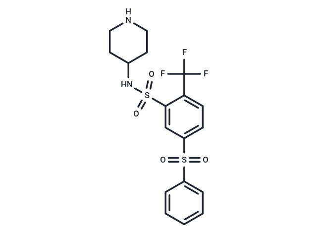
| Description | WAY 316606 is an inhibitor of the secreted protein sFRP-1, an endogenous antagonist of the secreted glycoprotein [Wnt]. |
| Targets & IC50 | SFRP1:0.5 μM |
| In vitro | The EC50 of WAY-316606 for Wnt-Luciferase Activity from U2-OS Cells is 0.65 μM[1]. WAY-316606 binds to the secreted frizzled-related protein (sFRP)-1 inhibitor with a KD of 0.08 μM and inhibits sFRP-1 with an EC50 of 0.65 μM. WAY-316606 also binds to sFRP-2, albeit over 10 times weaker with a KD of 1 μM. Using a fluorescence polarization binding assay that employs a fluorescent probe compound and purified human sFRP-1 protein in a competitive-binding format, the IC50 for WAY-316606 is 0.5 μM [2]. |
| In vivo | WAY-316606 effectively promotes bone formation, as demonstrated in neonatal murine calvarial assays, with the capability to increase total bone area by up to 60% in a dose-dependent manner, achieving an EC50 of approximately 1 nM. This compound exhibits favorable aqueous solubility, displays moderate to low inhibition of cytochrome P450 isozymes (3A4, 2D6, 2C9), and maintains good stability in both rat and human liver microsomes (t1/2>60 min for each species). Moreover, in female Sprague-Dawley rats, WAY-316606 shows high plasma clearance (77 mL/min/kg, surpassing hepatic blood flow) following a single intravenous bolus dose (2 mg/kg), leading to a swift reduction in plasma drug levels regardless of administration route [2]. |
| Kinase Assay | WAY-316606 binding to purified sFRP is determined by spectroscopy methods. The sFRP-1 or -2 stock solutions are diluted to 1 μM in a buffered solution and the initial fluorescence is measured. Increasing concentrations of WAY-316606 (0 to 50 μM) are added to the protein in the cuvette and incubated for 5 min prior to assessing fluorescence intensity using a Fluoromax-2 fluorometer. In control experiments, the DMSO (vehicle control)-matched buffer solution is used. Fluorescence spectra are scanned in the ratio mode (S/R, signal/reference) to compensate for variations in lamp output as a function of wavelength [2]. |
| Cell Research | U2OS bone cells are infected with recombinant adenovirus 5 (Ad5)?WNT3 at a multiplicity of infection (MOI) of 2, followed by infection with Ad5-sFRP-1 and Ad5-16xTCF-luciferase, each at an MOI of 10. Four hours after infection, the cells are frozen in sterile cryogenic vials at a cell density of 9×10⁶ cells/mL and stored in a ?150°C freezer. For the assay, a vial of frozen cells is thawed, and the cells are resuspended in plating medium [phenol red-free RPMI 1640 medium containing 5% fetal calf serum, 2 mM GlutaMAX-l, and 1% (v/v) penicillin-streptomycin] to a final cell density of 1.5×10⁵ cells/mL. The resuspended cells are then plated in 96-well tissue culture treated plates at a volume of 100 μL of cell suspension/well (i.e., 1.5×10⁴ cells/well). The plates are incubated at 37°C inside a 5% CO2/ 95% humidified air incubator for 5 h or until the cells have attached and started to spread. Prior to the addition of WAY-316606, the medium is replaced with 50 μL/well of phenol red-free RPMI 1640 containing 10% fetal calf serum, 2 mM GlutaMAX-l, and 1% (v/v) penicillin-streptomycin. WAY-316606, or vehicle (typically DMSO), diluted in phenol red-free RPMI 1640 containing 2 mM GlutaMAX-l, and l % (v/v) penicillin-streptomycin are then added to the wells in replicates of 4 wells/dilution and the plates are incubated at 37°C overnight. Dose?response experiments are performed with the compounds in 2-fold serial dilutions from 10000?4.9 nM. After the overnight incubation, the cells are washed twice with 150 uL/well of PBS w/o calcium or magnesium and lysed with 50 μL/well of 1× cell culture lysis reagent on a shaker at room temperature for 30 min. Aliquots of the cell lysates (30 μL) are transferred to 96-well luminometer plates, and the luciferase activity is measured in a MicroLumat PLUS luminometer using 100 μL/well of a luciferase substrate. |
| Molecular Weight | 448.48 |
| Formula | C18H19F3N2O4S2 |
| Cas No. | 915759-45-4 |
| Smiles | FC(F)(F)c1ccc(cc1S(=O)(=O)NC1CCNCC1)S(=O)(=O)c1ccccc1 |
| Relative Density. | 1.50 g/cm3 (Predicted) |
| Storage | Powder: -20°C for 3 years | In solvent: -80°C for 1 year Shipping with blue ice/Shipping at ambient temperature. | |||||||||||||||||||||||||||||||||||
| Solubility Information | DMSO: 125 mg/mL (278.72 mM), Sonication is recommended. | |||||||||||||||||||||||||||||||||||
| In Vivo Formulation | 10% DMSO+40% PEG300+5% Tween 80+45% Saline: 10 mg/mL (22.3 mM), Solution. 10% DMSO+90% Saline: < 10 mg/mL (22.3 mM), Lower concentrations may be soluble, but exact solubility limit is unknown. Please add the solvents sequentially, clarifying the solution as much as possible before adding the next one. Dissolve by heating and/or sonication if necessary. Working solution is recommended to be prepared and used immediately. The formulation provided above is for reference purposes only. In vivo formulations may vary and should be modified based on specific experimental conditions. | |||||||||||||||||||||||||||||||||||
Solution Preparation Table | ||||||||||||||||||||||||||||||||||||
DMSO
Note : The dilution table applies only to solid products. For liquid products, please calculate the stock solution based on the stated concentration and/or density. | ||||||||||||||||||||||||||||||||||||
 For example, if the intended dosage is 10 mg/kg for animals weighing 20 g , with a dosing volume of 100 μL per animal, and a total of 10 animals are to be administered, using a formulation of
  10% DMSO+ 40% PEG300+ 5% Tween 80+ 45% Saline/PBS/ddH2O , the resulting working solution concentration would be 2 mg/mL.
Dissolve 2 mg of the compound in 100 μL DMSO to obtain a stock solution at a concentration of 20 mg/mL . If the required concentration exceeds the compound's known solubility, please contact us for technical support before proceeding.
1) Add 100 μL of the DMSO stock solution to 400 µL PEG300 and mix thoroughly until the solution becomes clear.
2) Add 50 µL Tween 80 and mix well until fully clarified.
3) Add 450 µL Saline,PBS or ddH2O and mix thoroughly until a homogeneous solution is obtained.
| Size | Quantity | Unit Price | Amount | Operation |
|---|
 Hello! How can I help you today?
 Copyright © 2015-2026 TargetMol Chemicals Inc. All Rights Reserved.
