
Shopping Cart
Remove All Your shopping cart is currently empty


Synonyms:

| Pack Size | Price | USA Stock | Global Stock | Quantity |
|---|---|---|---|---|
| 50 µg | $44 | In Stock | In Stock | |
| 100 µg | $73 | In Stock | In Stock | |
| 500 µg | $250 | In Stock | In Stock | |
| 1 mg | $376 | In Stock | In Stock | |
| 5 mg | $856 | In Stock | In Stock | |
| 10 mg | $1,130 | In Stock | In Stock |
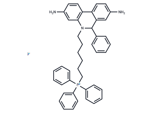
| Description | MitoSOX Red is a fluorescent dye serving as a mitochondria-targeted fluorescent probe for superoxide, with cell membrane permeability and specificity. Upon oxidation, MitoSOX Red subsequently binds to nucleic acids within mitochondria/nuclei, producing intense red fluorescence, and is employed for detecting mitochondrial superoxide in live cells. |
| In vitro | I. Solution Preparation 1. MitoSOX Red Stock Solution Preparation: Dissolve MitoSOX Red in DMSO, typically at a concentration of 1-10 mM, which can be optimized based on experimental conditions. 2. Working Solution Preparation: Dilute the MitoSOX Red stock solution to a concentration suitable for the experiment. The common final concentration range is 1 to 10 µM, but this should be optimized according to cell type and experimental conditions. 3. Control Reagents: Use superoxide dismutase (SOD) as a negative control, as SOD scavenges superoxide and prevents the oxidation of MitoSOX Red. II. Cell Labeling 1. Cell Culture: Seed cells into appropriate culture vessels (such as culture dishes or coverslips) and culture for 24-48 hours until cells are fully attached and grown. 2. MitoSOX Red Staining: Add working concentration of MitoSOX Red to the culture medium. Incubate at 37°C for 10 to 30 minutes; staining time should be optimized according to cell type. 3. Washing: After incubation, thoroughly wash cells with PBS to remove excess MitoSOX Red. 4. Fluorescence Detection: 1) Excitation and Emission Wavelengths: The excitation wavelength of MitoSOX Red is 510 nm, and the emission wavelength is 580 nm. Use a fluorescence microscope or flow cytometer equipped with appropriate filters to detect red fluorescence. 2) Superoxide Detection: The intensity of red fluorescence is proportional to the superoxide level in mitochondria. Higher fluorescence intensity indicates higher superoxide levels, while low fluorescence indicates lower superoxide levels. 5. Experimental Controls: 1) Positive Control: Use known oxidative stress inducers, such as Paraquat or Rotenone, to elevate intracellular superoxide levels, thereby increasing MitoSOX Red fluorescence. 2) Negative Control: Add Mito-TEMPO to prevent superoxide from oxidizing MitoSOX Red, thereby reducing or eliminating fluorescence. 6. Quantitative Analysis: For quantitative analysis, flow cytometry can be used to measure fluorescence intensity of individual cells, which helps analyze superoxide levels in large cell populations. Qualitative assessment can also be performed through image analysis. |
| Cell Research | I. Solution preparation 1. MitoSOX Red stock solution preparation: MitoSOX Red is dissolved in DMSO, usually at a concentration of 1-10mM, which can be optimized according to experimental conditions. 2. Working solution preparation: Dilute the MitoSOX Red stock solution to a concentration suitable for the experiment. Common final concentrations range from 1 to 10 µM, but should be optimized according to cell type and experimental conditions. 3. Control reagent: Use superoxide dismutase (SOD) as a negative control because SOD will scavenge superoxide and prevent oxidation of MitoSOX Red. II. Cell labeling 1. Cultivate cells: Inoculate cells into appropriate culture vessels (such as culture dishes or coverslips) and culture for 24-48 hours until cells are fully attached and growing. 2. MitoSOX Red staining: Add the working concentration of MitoSOX Red to the culture medium. Incubate at 37°C for 10 to 30 minutes. Staining time should be optimized based on cell type. 3. Washing: After incubation, wash cells thoroughly with PBS to remove excess MitoSOX Red. 4. Fluorescence detection: 1) Excitation and emission wavelengths: MitoSOX Red has an excitation wavelength of 510 nm and an emission wavelength of 580 nm. Use a fluorescence microscope or flow cytometer equipped with appropriate filters to detect red fluorescence. 2) Superoxide detection: The intensity of red fluorescence is proportional to the level of superoxide in mitochondria. Higher fluorescence intensity indicates higher superoxide levels, while lower fluorescence indicates lower superoxide levels. 5. Experimental controls: 1) Positive control: Use known oxidative stress inducers, such as Paraquat or Rotenone, to elevate superoxide levels in cells, thereby increasing the fluorescence of MitoSOX Red. 2) Negative control: Add Mito-TEMPO to prevent superoxide from oxidizing MitoSOX Red, thereby reducing or eliminating fluorescence. 6. Quantitative analysis: For quantitative analysis, flow cytometry can be used to measure the fluorescence intensity of individual cells, which helps to analyze superoxide levels in a large number of cells. Qualitative assessment can also be performed by image analysis. Notes: 1. MitoSOX Red is light-sensitive, so it should be avoided from exposure to light during storage and operation. 2. This probe specifically targets mitochondria and will only be oxidized by superoxide, not other ROS or RNS, thus ensuring the specificity of the detection. 3. During the incubation process, ensure that the cells are not overly stressed or damaged to avoid causing artifacts or nonspecific fluorescence. The above information is based on published literature. Experimental procedures should be appropriately modified to meet specific research demands. |
| Molecular Weight | 759.7 |
| Formula | C43H43IN3P |
| Cas No. | 1003197-00-9 |
| Smiles | [I-].NC=1C=CC=2C3=CC=C(N)C=C3C(C=4C=CC=CC4)N(C2C1)CCCCCC[P+](C=5C=CC=CC5)(C=6C=CC=CC6)C=7C=CC=CC7 |
| Storage | Keep away from direct sunlight,Store at low temperature,Keep away from moisture Powder: -20°C for 3 years | In solvent: -80°C for 1 year Shipping with blue ice/Shipping at ambient temperature. | |||||||||||||||||||||||||||||||||||
| Solubility Information | DMSO: 101 mg/mL (132.95 mM), Sonication is recommended. | |||||||||||||||||||||||||||||||||||
| In Vivo Formulation | 10% DMSO+40% PEG300+5% Tween-80+45% Saline: 3.3 mg/mL (4.34 mM), Sonication is recommended. Please add the solvents sequentially, clarifying the solution as much as possible before adding the next one. Dissolve by heating and/or sonication if necessary. Working solution is recommended to be prepared and used immediately. The formulation provided above is for reference purposes only. In vivo formulations may vary and should be modified based on specific experimental conditions. | |||||||||||||||||||||||||||||||||||
Solution Preparation Table | ||||||||||||||||||||||||||||||||||||
DMSO
Note : The dilution table applies only to solid products. For liquid products, please calculate the stock solution based on the stated concentration and/or density. | ||||||||||||||||||||||||||||||||||||
 For example, if the intended dosage is 10 mg/kg for animals weighing 20 g , with a dosing volume of 100 μL per animal, and a total of 10 animals are to be administered, using a formulation of
  10% DMSO+ 40% PEG300+ 5% Tween 80+ 45% Saline/PBS/ddH2O , the resulting working solution concentration would be 2 mg/mL.
Dissolve 2 mg of the compound in 100 μL DMSO to obtain a stock solution at a concentration of 20 mg/mL . If the required concentration exceeds the compound's known solubility, please contact us for technical support before proceeding.
1) Add 100 μL of the DMSO stock solution to 400 µL PEG300 and mix thoroughly until the solution becomes clear.
2) Add 50 µL Tween 80 and mix well until fully clarified.
3) Add 450 µL Saline,PBS or ddH2O and mix thoroughly until a homogeneous solution is obtained.
| Size | Quantity | Unit Price | Amount | Operation |
|---|
 Hello! How can I help you today?
 Copyright © 2015-2026 TargetMol Chemicals Inc. All Rights Reserved.
