 Your shopping cart is currently empty

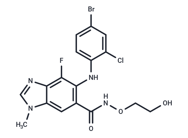
Selumetinib (AZD6244) is a MEK1/2 inhibitor that inhibits MEK1 (IC50=14 nM) with potent selectivity and is non-ATP-competitive. Selumetinib has antitumor activity and is used for the treatment of neurofibromatosis type 1 (NF1).

| Pack Size | Price | USA Warehouse | Global Warehouse | Quantity |
|---|---|---|---|---|
| 25 mg | $47 | In Stock | In Stock | |
| 50 mg | $63 | In Stock | In Stock | |
| 100 mg | $81 | In Stock | In Stock | |
| 200 mg | $126 | In Stock | In Stock | |
| 500 mg | $198 | In Stock | In Stock | |
| 1 g | $297 | In Stock | In Stock |
| Description | Selumetinib (AZD6244) is a MEK1/2 inhibitor that inhibits MEK1 (IC50=14 nM) with potent selectivity and is non-ATP-competitive. Selumetinib has antitumor activity and is used for the treatment of neurofibromatosis type 1 (NF1). |
| Targets&IC50 | ERK1/2 phosphorylation:10.3 nM (Malme-3M cells), MEK:12 nM, MEK1:14 nM (cell free) |
| In vitro | METHODS: TNBC cell lines MDA-MB-231, MDA-MB-468, SUM149, SUM190, KPL-4, and MDA-IBC-3 were treated with Selumetinib (0-100 µM) for 72 h, and cell viability was measured by WST-1 assay. RESULTS: In MDA-MB-468, SUM190, KPL-4 and MDA-IBC-3 cells, the IC50 was above 20 µM, and in MDA-MAB-231 and SUM149 cells, the IC50 values were 8.6 µM and 10 µM, respectively. [1] METHODS: Breast cancer cells HCC-1937 and MDA-MB-231 were treated with Selumetinib (1-50 µM) for 24 h, and the cell cycle was detected by Flow cytometry. RESULTS: Selumetinib triggered apoptosis and G1 phase block in a dose-dependent manner. [2] |
| In vivo | METHODS: To assay antitumor activity in vivo, Selumetinib (50 mg/kg, 0.5% hydroxypropyl methyl cellulose and 0.1% Tween 80) was administered by gavage to athymic nu/nu mice bearing MDA-MB-231-LM2 xenografts five times per week for three weeks. for three weeks. RESULTS: In mice treated with Selumetinib, lung metastases were inhibited and tumor cells reversed from a mesenchymal phenotype to an epithelial phenotype. [1] |
| Kinase Assay | NH2-terminal hexahistidine tagged, constitutively active MEK1 was expressed in baculovirus-infected Hi5 insect cells and purified by immobilized metal affinity chromatography, ion exchange, and gel filtration. The activity of MEK1 was assessed by measuring the incorporation of [γ-33P]phosphate from [γ-33P]ATP onto ERK2. The assay was carried out in a 96-well polypropylene plate with an incubation mixture (100 μL) composed of 25 mmol/L HEPES (pH 7.4), 10 mmol/L MgCl2, 5 mmol/L β-glycerolphosphate, 100 μmol/L sodium orthovanadate, 5 mmol/L DTT, 5 nmol/L MEK1, 1 μmol/L ERK2, and 0 to 80 nmol/L compound (final concentration of 1% DMSO). The reactions were initiated by the addition of 10 μmol/L ATP (with 0.5 μC k[γ-33P]ATP/well) and incubated at room temperature for 45 min. An equal volume of 25% trichloracetic acid was added to stop the reaction and precipitate the proteins. Precipitated proteins were trapped onto glass fiber B filter plates, excess labeled ATP was washed off with 0.5% phosphoric acid, and radioactivity was counted in a liquid scintillation counter. ATP dependence was determined by varying the amount of ATP in the reaction mixture. The data were globally fitted using SigmaPlot. Values were calculated using the following equation for noncompetitive inhibition: v = [Vmax × S / (1 + I / Ki)] / (Km + S) [1]. |
| Cell Research | Primary HCC cells were plated at a density of 2.0 × 10^4 per well in growth medium. After 48 h in growth medium, the cell monolayer was rinsed twice with MEM. Cells were treated with various concentrations of AZD6244 (0, 0.5, 1.0, 2.0, 3.0, and 4.0 μmol/L) for 24 or 48 h. Cell viability was determined by the MTT assay. Cell proliferation was assayed using a bromodeoxyuridine kit as described by the manufacturer. Experiments were repeated at least thrice, and the data were expressed as mean ± SE [2]. |
| Animal Research | HT-29 human colon carcinoma or BxPC3 human pancreatic tumor fragments were implanted s.c. in the flank of nude mice and allowed to grow to 100 to 150 mg. Mice (n = 10 per group) were randomized to treatment groups to receive vehicle (10 mL/kg and 10% ethanol/10% cremophor EL/80% D5W) or ARRY-142886 (10, 25, 50, or 100 mg/kg, oral, BID) on days 1 to 21. Tumors [(W^2 × L) / L] were measured twice weekly. Tumor growth inhibition was calculated as 1 ? (tumor sizetreated / tumor sizevehicle) on each measurement day. Four hours after the last dose on day 21, three mice per group were euthanized to evaluate pharmacokinetic/pharmacodynamic responses. Tumors were excised and flash frozen. Homogenates were analyzed for phospho-ERK1/2 and ERK1/2 expression by Western blotting as described above. For the HT-29 study, monitoring of tumor regrowth was continued for the remaining seven mice per group until tumors reached 1,000 mm^3, when mice would be sacrificed. There were two BxPC3 tumor xenograft studies. For the first study, one group of mice was treated with the clinical standard of care, gemcitabine, at 160 mg/kg, i.p., every 3rd day for a total of four doses. This dose was determined to be the maximum tolerated dose for gemcitabine in the BxPC3 model on this dosing schedule. To evaluate whether previously treated tumors would be refractory to a second cycle of treatment, a second BxPC3 xenograft study was carried out. Mice were treated with vehicle or ARRY-142886 at 25 or 50 mg/kg, BID, for 21 days. Treatment was stopped and tumors were allowed to grow for an additional 7 days before treatment resumed for another 21-day cycle [1]. |
| Synonyms | AZD6244, ARRY-142886 |
| Molecular Weight | 457.68 |
| Formula | C17H15BrClFN4O3 |
| Cas No. | 606143-52-6 |
| Smiles | CN1C=NC2=C(F)C(NC3=CC=C(Br)C=C3Cl)=C(C=C12)C(=O)NOCCO |
| Relative Density. | 1.69 |
| Storage | store at low temperature | In solvent: -80°C for 1 year | Shipping with blue ice/Shipping at ambient temperature. | |||||||||||||||||||||||||||||||||||
| Solubility Information | DMSO: 81.7 mg/mL (178.51 mM), Sonication is recommended. Ethanol: < 1 mg/mL (insoluble or slightly soluble) H2O: < 1 mg/mL (insoluble or slightly soluble) | |||||||||||||||||||||||||||||||||||
| In Vivo Formulation | 10% DMSO+40% PEG300+5% Tween 80+45% Saline: 2 mg/mL (4.37 mM), Sonication is recommended. Please add the solvents sequentially, clarifying the solution as much as possible before adding the next one. Dissolve by heating and/or sonication if necessary. Working solution is recommended to be prepared and used immediately. The formulation provided above is for reference purposes only. In vivo formulations may vary and should be modified based on specific experimental conditions. | |||||||||||||||||||||||||||||||||||
Solution Preparation Table | ||||||||||||||||||||||||||||||||||||
DMSO
| ||||||||||||||||||||||||||||||||||||
Dissolve 2 mg of the compound in 100 μL DMSO![]() to obtain a stock solution at a concentration of 20 mg/mL . If the required concentration exceeds the compound's known solubility, please contact us for technical support before proceeding.
to obtain a stock solution at a concentration of 20 mg/mL . If the required concentration exceeds the compound's known solubility, please contact us for technical support before proceeding.
1) Add 100 μL of the DMSO![]() stock solution to 400 μL PEG300
stock solution to 400 μL PEG300![]() and mix thoroughly until the solution becomes clear.
and mix thoroughly until the solution becomes clear.
2) Add 50 μL Tween 80 and mix well until fully clarified.
3) Add 450 μL Saline,PBS or ddH2O![]() and mix thoroughly until a homogeneous solution is obtained.
and mix thoroughly until a homogeneous solution is obtained.
| Size | Quantity | Unit Price | Amount | Operation |
|---|

Copyright © 2015-2026 TargetMol Chemicals Inc. All Rights Reserved.