
Shopping Cart
Remove All Your shopping cart is currently empty

Synonyms: Protein tyrosine kinase ERK 2, Protein kinase mitogen activated 3, Protein kinase mitogen activated 2, Protein kinase mitogen activated 1, PRKM3, PRKM2, PRKM1, PRKM 3, PRKM 2, PRKM 1, p44MAPK, p44ERK1, p44 MAPK, p44 ERK1, p42-MAPK, p42MAPK, p42 MAPK, p41mapk, p41, p40, p38, MK03, MK01, Mitogen activated protein kinase 3, Mitogen activated protein kinase 2, Mitogen activated protein kinase 1, Microtubule associated protein 2 kinase, MGC20180, MAPK3, MAPK2, MAPK1, MAPK 3, MAPK 2, MAPK 1, MAP kinase isoform p44, MAP kinase isoform p42, MAP kinase 2, MAP kinase 1, Insulin stimulated MAP2 kinase, HUMKER1A, HS44KDAP, Extracellular signal-regulated kinase 2, Extracellular signal regulated kinase 2, Extracellular signal regulated kinase 1, ERT2, ERT1, ERK-2, ERK2, ERK1, ERK 2, ERK 1/2, ERK 1
Anti-ERK1/2 Polyclonal Antibody
| Pack Size | Price | USA Stock | Global Stock | Quantity |
|---|---|---|---|---|
| 50 µL | $222 | 7-10 days | 7-10 days | |
| 100 µL | $373 | 7-10 days | 7-10 days | |
| 200 µL | $528 | 7-10 days | 7-10 days |
| Description | Anti-ERK1/2 Polyclonal Antibody is a Rabbit antibody targeting ERK1/2. Anti-ERK1/2 Polyclonal Antibody can be used in FCM,IF,IHC-Fr,IHC-P,WB. |
| Synonyms | Protein tyrosine kinase ERK 2, Protein kinase mitogen activated 3, Protein kinase mitogen activated 2, Protein kinase mitogen activated 1, PRKM3, PRKM2, PRKM1, PRKM 3, PRKM 2, PRKM 1, p44MAPK, p44ERK1, p44 MAPK, p44 ERK1, p42-MAPK, p42MAPK, p42 MAPK, p41mapk, p41, p40, p38, MK03, MK01, Mitogen activated protein kinase 3, Mitogen activated protein kinase 2, Mitogen activated protein kinase 1, Microtubule associated protein 2 kinase, MGC20180, MAPK3, MAPK2, MAPK1, MAPK 3, MAPK 2, MAPK 1, MAP kinase isoform p44, MAP kinase isoform p42, MAP kinase 2, MAP kinase 1, Insulin stimulated MAP2 kinase, HUMKER1A, HS44KDAP, Extracellular signal-regulated kinase 2, Extracellular signal regulated kinase 2, Extracellular signal regulated kinase 1, ERT2, ERT1, ERK-2, ERK2, ERK1, ERK 2, ERK 1/2, ERK 1 |
| Ig Type | IgG |
| Reactivity | Human,Mouse,Rat (predicted:Chicken,Dog,Pig,Cow,Horse,Rabbit,Sheep,Goat) |
| Verified Activity | 1. Paraformaldehyde-fixed, paraffin embedded (Mouse brain); Antigen retrieval by boiling in sodium citrate buffer (pH6.0) for 15 min; Block endogenous peroxidase by 3% hydrogen peroxide for 20 min; Blocking buffer (normal goat serum) at 37°C for 30 min; Antibody incubation with (ERK1 + ERK2) Polyclonal Antibody, Unconjugated (TMAB-00633) at 1:400 overnight at 4°C, followed by operating according to SP Kit (Rabbit) instructionsand DAB staining. 2. Sample: Brain (Rat) Lysate at 30 μg Heart (Rat) lysate at 30 μg Primary: Anti-ERK2/MAPK1 (TMAB-00633) at 1/200 dilution Secondary: HRP conjugated Goat-Anti-rabbit IgG (secondary antibody) at 1/3000 dilution Predicted band size: 42 kDa Observed band size: 42 kDa 3. Tissue/cell: human lung carcinoma; 4% Paraformaldehyde-fixed and paraffin-embedded; Antigen retrieval: citrate buffer (0.01M, pH6.0), Boiling bathing for 15 min; Block endogenous peroxidase by 3% Hydrogen peroxide for 30 min; Blocking buffer (normal goat serum) at 37°C for 20 min; Incubation: Anti-ERK2/MAPK1 Polyclonal Antibody, Unconjugated (TMAB-00633) 1:200, overnight at 4°C, followed by conjugation to the secondary antibody and DAb staining. 4. Blank control: Hep G2 cells (blue). Primary Antibody: Rabbit Anti-ERK1 + ERK2 antibody (TMAB-00633), Dilution: 1 μg in 100 μL 1X PBS containing 0.5% BSA; Isotype Control Antibody: Rabbit Igg (orange),used under the same conditions); Secondary Antibody: Goat anti-rabbit IgG-Pe (white blue), Dilution: 1:200 in 1 X PBS containing 0.5% BSA. Protocol The cells were fixed with 2% paraformaldehyde (10 min), then permeabilized with 90% ice-cold methanol for 30 min on ice. Primary antibody (TMAB-00633, 1 μg/1x10^6 cells) were incubated for 30 min on the ice, followed by 1 X PBS containing 0.5% BSA + 10% goat serum (15 min) to block non-specific protein-protein interactions. Then the Goat Anti-rabbit IgG/PE antibody was added into the blocking buffer mentioned above to react with the primary antibody at 1/200 dilution for 30 min on ice. 5. Sample: Lane 1: A431 (Human) Cell Lysate at 30 μg Lane 2: MCF-7 (Human) Cell Lysate at 30 μg Lane 3: Huvec (Human) Cell Lysate at 30 μg Primary: Anti-ERK1 + ERK2 (TMAB-00633) at 1/1000 dilution Secondary: IRDye800CW Goat Anti-Rabbit IgG at 1/20000 dilution Predicted band size: 44/42 kDa Observed band size: 42/40 kDa 6. Sample: Lane 1: Cerebrum (Mouse) Lysate at 40 μg Lane 2: Cerebrum (Rat) Lysate at 40 μg Lane 3: LympHnode (Mouse) Lysate at 40 μg Lane 4: LympHnode (Rat) Lysate at 40 μg Primary: Anti-ERK1 + ERK2 (TMAB-00633) at 1/1000 dilution Secondary: IRDye800CW Goat Anti-Rabbit IgG at 1/20000 dilution Predicted band size: 44/42 kDa Observed band size: 40 kDa 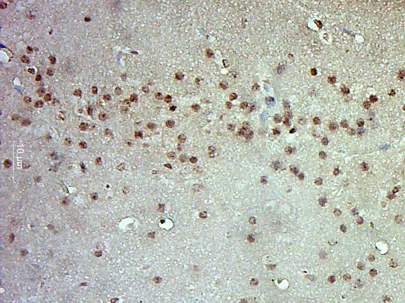 , ,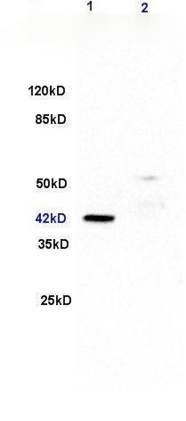 , ,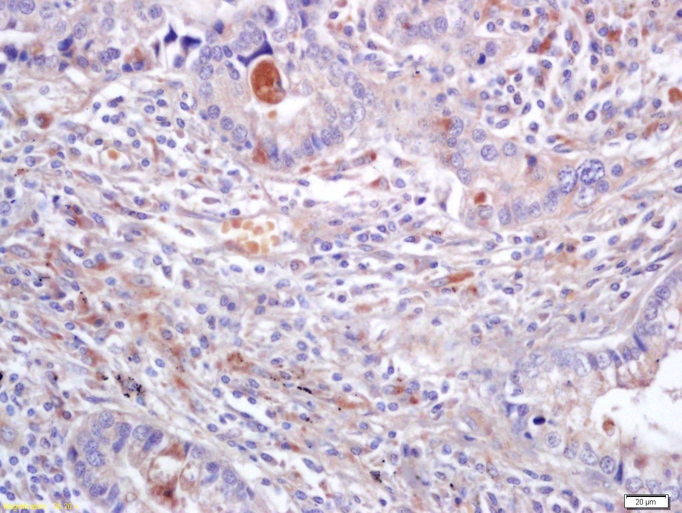 , ,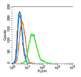 , ,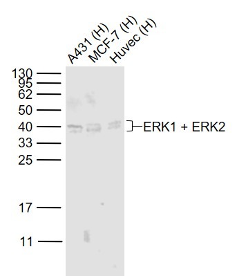 , ,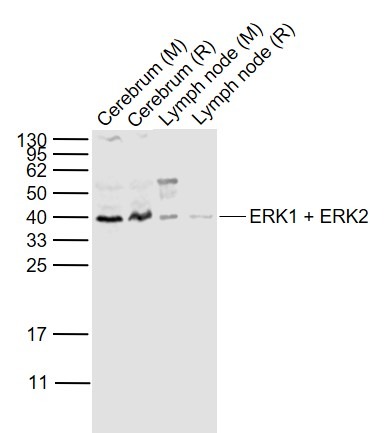 |
| Application | |
| Recommended Dose | WB: 1:500-2000; IHC-P: 1:100-500; IHC-Fr: 1:100-500; IF: 1:100-500; FCM: 1μg/Test |
| Antibody Type | Polyclonal |
| Host Species | Rabbit |
| Subcellular Localization | Cytoplasm, cytoskeleton, spindle. Nucleus. Cytoplasm, cytoskeleton, centrosome. Cytoplasm. Note=Associated with the spindle duringprometaphase and metaphase. PEA15-binding andphosphorylated DAPK1 promote its cytoplasmic retention.Phosphorylation at Ser-244 and Ser-246 as well asautophosphorylation at Thr-188 promote nuclear localization. |
| Tissue Specificity | Widely expressed. |
| Construction | Polyclonal Antibody |
| Purification | Protein A purified |
| Appearance | Liquid |
| Formulation | 0.01M TBS (pH7.4) with 1% BSA, 0.02% Proclin300 and 50% Glycerol. |
| Concentration | 1 mg/mL |
| Research Background | The protein encoded by this gene is a member of the MAPkinase family. MAP kinases, also known as extracellularsignal-regulated kinases (ERKs), act in a signaling cascade thatregulates various cellular processes such as proliferation,differentiation, and cell cycle progression in response to avariety of extracellular signals. This kinase is activated byupstream kinases, resulting in its translocation to the nucleuswhere it phosphorylates nuclear targets. Alternatively splicedtranscript variants encoding different protein isoforms have beendescribed. [provided by RefSeq, Jul 2008]. |
| Immunogen | KLH conjugated synthetic peptide: human ERK2 |
| Antigen Species | Human |
| Gene Name | MAPK3 |
| Gene ID | |
| Protein Name | Mitogen-activated protein kinase 3 |
| Uniprot ID | |
| Biology Area | Tangles & Tau,MAPK Pathway,Cytoplasmic |
| Function | Serine/threonine kinase which acts as an essentialcomponent of the MAP kinase signal transduction pathway. MAPK1/ERK2and MAPK3/ERK1 are the 2 MAPKs which play an important role in theMAPK/ERK cascade. They participate also in a signaling cascadeinitiated by activated KIT and KITLG/SCF. Depending on the cellularcontext, the MAPK/ERK cascade mediates diverse biological functionssuch as cell growth, adhesion, survival and differentiation throughthe regulation of transcription, translation, cytoskeletalrearrangements. The MAPK/ERK cascade plays also a role ininitiation and regulation of meiosis, mitosis, and postmitoticfunctions in differentiated cells by phosphorylating a number oftranscription factors. About 160 substrates have already beendiscovered for ERKs. Many of these substrates are localized in thenucleus, and seem to participate in the regulation of transcriptionupon stimulation. However, other substrates are found in thecytosol as well as in other cellular organelles, and those areresponsible for processes such as translation, mitosis andapoptosis. Moreover, the MAPK/ERK cascade is also involved in theregulation of the endosomal dynamics, including lysosome processingand endosome cycling through the perinuclear recycling compartment(PNRC); as well as in the fragmentation of the Golgi apparatusduring mitosis. The substrates include transcription factors (suchas ATF2, BCL6, ELK1, ERF, FOS, HSF4 or SPZ1), cytoskeletal elements(such as CANX, CTTN, GJA1, MAP2, MAPT, PXN, SORBS3 or STMN1),regulators of apoptosis (such as BAD, BTG2, CASP9, DAPK1, IER3,MCL1 or PPARG), regulators of translation (such as EIF4EBP1) and avariety of other signaling-related molecules (like ARHGEF2, DCC,FRS2 or GRB10). Protein kinases (such as RAF1, RPS6KA1/RSK1,RPS6KA3/RSK2, RPS6KA2/RSK3, RPS6KA6/RSK4, SYK, MKNK1/MNK1,MKNK2/MNK2, RPS6KA5/MSK1, RPS6KA4/MSK2, MAPKAPK3 or MAPKAPK5) andphosphatases (such as DUSP1, DUSP4, DUSP6 or DUSP16) are othersubstrates which enable the propagation the MAPK/ERK signal toadditional cytosolic and nuclear targets, thereby extending thespecificity of the cascade. Mediates phosphorylation of TPR inrespons to EGF stimulation. May play a role in the spindle assemblycheckpoint. Phosphorylates PML and promotes its interaction withPIN1, leading to PML degradation (By similarity). Acts as a transcriptional repressor. Binds to a[GC]AAA[GC] consensus sequence. Repress the expression ofinterferon gamma-induced genes. Seems to bind to the promoter ofCCL5, DMP1, IFIH1, IFITM1, IRF7, IRF9, LAMP3, OAS1, OAS2, OAS3 andSTAT1. Transcriptional activity is independent of kinase activity. |
| Molecular Weight | Theoretical: 42 kDa. Actual: 40-44 kDa. |
| Stability & Storage | Store at -20°C or -80°C for 12 months. Avoid repeated freeze-thaw cycles. |
| Transport | Shipping with blue ice. |
| Size | Quantity | Unit Price | Amount | Operation |
|---|
 Hello! How can I help you today?
 Copyright © 2015-2026 TargetMol Chemicals Inc. All Rights Reserved.
