
Shopping Cart
Remove All Your shopping cart is currently empty

Synonyms:
| Pack Size | Price | USA Stock | Global Stock | Quantity |
|---|---|---|---|---|
| 50 mg | $40 | - | In Stock | |
| 100 mg | $56 | - | In Stock | |
| 1 mL x 10 mM (in DMSO) | $29 | - | In Stock |
| Description | Murrayafoline A is a natural carbazole alkaloid found primarily in plants of the genera Murraya and Glycosmis. Murrayafoline A directly targets Specific Protein 1 (Sp1), thereby inhibiting the NF-κB and MAPK signaling pathways. Murrayafoline A attenuates the Wnt/β-catenin pathway by promoting the degradation of intracellular β-catenin. Murrayafoline A induces G0/G1 phase arrest in platelet-derived growth factor (PDGF)-stimulated vascular smooth muscle cells. Murrayafoline A enhances contractility and L-type calcium currents in rat ventricular myocytes by activating protein kinase C. Murrayafoline A inhibits LPS-induced neuroinflammation in vivo. Murrayafoline A can be used in research on inflammation, vascular complications, and colorectal cancer. |
| In vitro | Methods: BV-2 cells were treated with Murrayafoline A (20 μM) and LPS (1 μg/mL) for 24 hours. TNF-α and IL-6 levels in the cell culture supernatant were measured by ELISA, and IL-1β and iNOS mRNA expression levels were assessed by qPCR. Results: Murrayafoline A significantly inhibited LPS-induced TNF-α and IL-6 release, and significantly suppressed LPS-induced upregulation of IL-1β and iNOS mRNA. [1] Methods: Rat aortic VSMCs were pretreated with Murrayafoline A (1, 3, 5 μM) for 24 h, followed by treatment with PDGF-BB (50 ng/mL) for 24 h. Cells were stained with PI, analyzed by flow cytometry (FACS Calibur) using ModFit LT for cell cycle analysis, and Western blot was performed to detect the expression of cyclin D1, cyclin E, CDK2, CDK4, and PCNA. Results: Murrayafoline A significantly inhibited PDGF-BB-stimulated vascular smooth muscle cell proliferation and DNA synthesis, induced G0/G1 phase arrest, and simultaneously suppressed the expression of cyclin D1, cyclin E, CDK2, CDK4, and PCNA. [2] Methods: HEK293 reporter cells (TOPFlash) were treated with Murrayafoline A (2.5, 5, 10, 20 μM) and Wnt3a-CM for 15 hours. Cytoplasmic β-catenin protein levels were assessed by Western blot, and β-catenin mRNA expression was detected by semi-quantitative RT-PCR. Results: Murrayafoline A concentration-dependently reduced the Wnt3a-CM-induced increase in cytoplasmic β-catenin levels but had no effect on β-catenin mRNA levels. [3] Methods: Murrayafoline A (25 μM) was added to rat ventricular myocytes, and membrane potential changes were continuously recorded using the patch-clamp technique. Results: Murrayafoline A increased cell shortening in rat ventricular myocytes, enhanced L-type Ca²⁺ currents, and promoted PKC phosphorylation. [4] Methods: Murrayafoline A (0.01–40 μg/mL) was added to human hepatocellular carcinoma cells (HepG2) and treated for 24, 48, 72, and 96 hours; cell viability was assessed using the MTT assay. Results: Murrayafoline A effectively killed hepatocellular carcinoma cells, with an IC₅₀ of approximately 7 μM.[5] |
| In vivo | Methods: To investigate the effects of Murrayafoline A on LPS-induced neuronal damage, BALB/c mice were administered Murrayafoline A (25 mg/kg) intraperitoneally once daily for 3 consecutive days. Two hours after the final dose, LPS (5 mg/kg) was administered intraperitoneally, and the mice were euthanized 6 hours later. Results: Murrayafoline A inhibited LPS-induced microglial activation, increased the number of Nissl bodies, and attenuated LPS-induced neuronal damage. [1] |
| Molecular Weight | 211.26 |
| Formula | C14H13NO |
| Cas No. | 4532-33-6 |
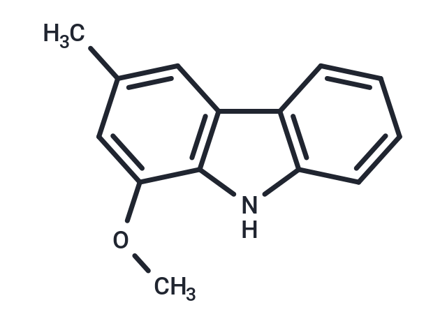
| Smiles | O(C1=CC(=CC=2C=3C=CC=CC3NC12)C)C |
| Storage | Powder: -20°C for 3 years | In solvent: -80°C for 1 year Shipping with blue ice/Shipping at ambient temperature. |
| Solubility Information | DMSO: 80.00 mg/mL (378.68 mM), Sonication is recommended. H2O: 0.08 mg/mL (0.38 mM), Sonication is recommended. |
 For example, if the intended dosage is 10 mg/kg for animals weighing 20 g , with a dosing volume of 100 μL per animal, and a total of 10 animals are to be administered, using a formulation of
  10% DMSO+ 40% PEG300+ 5% Tween 80+ 45% Saline/PBS/ddH2O , the resulting working solution concentration would be 2 mg/mL.
Dissolve 2 mg of the compound in 100 μL DMSO to obtain a stock solution at a concentration of 20 mg/mL . If the required concentration exceeds the compound's known solubility, please contact us for technical support before proceeding.
1) Add 100 μL of the DMSO stock solution to 400 µL PEG300 and mix thoroughly until the solution becomes clear.
2) Add 50 µL Tween 80 and mix well until fully clarified.
3) Add 450 µL Saline,PBS or ddH2O and mix thoroughly until a homogeneous solution is obtained.
| Size | Quantity | Unit Price | Amount | Operation |
|---|
 Hello! How can I help you today?
 Copyright © 2015-2026 TargetMol Chemicals Inc. All Rights Reserved.
