
Shopping Cart
Remove All Your shopping cart is currently empty


Synonyms:

| Pack Size | Price | USA Stock | Global Stock | Quantity |
|---|---|---|---|---|
| 1 mg | $59 | In Stock | In Stock | |
| 5 mg | $143 | In Stock | In Stock | |
| 10 mg | $228 | In Stock | In Stock | |
| 25 mg | $389 | In Stock | In Stock | |
| 50 mg | $579 | In Stock | In Stock | |
| 100 mg | $868 | In Stock | In Stock | |
| 1 mL x 10 mM (in DMSO) | $186 | In Stock | In Stock |
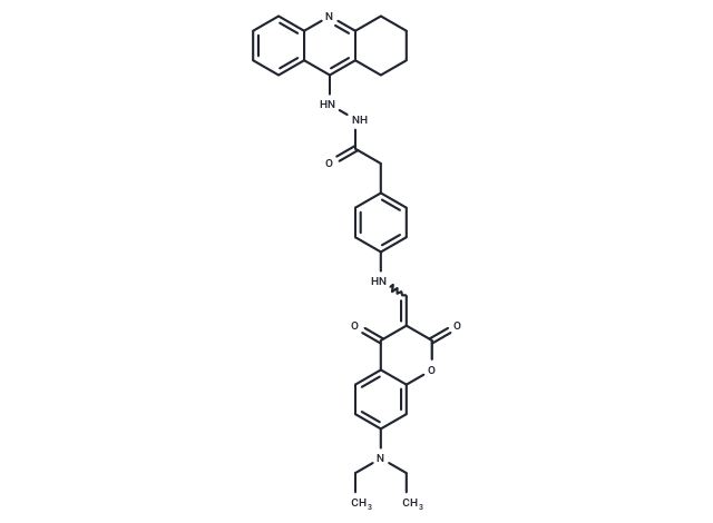
| Description | PE 154 is a potent fluorescent inhibitor of acetylcholinesterase (AChE) and butyrylcholinesterase (BChE) with IC50s of 280 pM and 16 nM respectively. PE 154 is commonly used to label β-amyloid plaques in histochemical analysis. |
| Targets & IC50 | BChE:16 nM, AChE:280 pM |
| Cell Research | Instructions I. Preparation of reagents Configuration of mother solution and working solution: Usually, the stock solution with a concentration of 1 mM can be diluted to obtain the required working concentration (e.g., 0.1–10 μM). The specific concentration is adjusted according to the experimental requirements and the characteristics of the target tissue. II. Labeling tissues and samples 1. Tissue preparation: When using PE 154 for tissue staining, you first need to prepare tissue sections. For mice or other animal models, brain tissue that has been properly fixed (such as using formaldehyde fixation) is usually selected. 2. Staining process: 1) Place tissue sections or cell samples in a staining solution containing PE 154 solution. The concentration of the staining solution is usually between 1 μM and 10 μM, which is adjusted according to experimental requirements. 2) The sample should be incubated at room temperature for 30 minutes to 1 hour. The incubation time depends on the type of sample and the specific requirements of the experiment. 3. Washing: After incubation, gently wash the sections with PBS or an appropriate buffer to remove unbound dye to reduce background signals. 3. Fluorescence imaging 1. PE 154 is a fluorescent marker, so it can be imaged by fluorescence microscope, confocal microscope and other equipment. 2. Its excitation wavelength is usually 470 nm and its emission wavelength is 520 nm. 3. Clear fluorescence signals can be observed under the microscope for the stained samples, especially when β-amyloid plaques are labeled, the plaque area will show strong fluorescence. 4. Data analysis 1. For the analysis of β-amyloid plaques, the fluorescence intensity of the sample can be directly observed by fluorescence microscopy, and the number, size and distribution of plaques can be quantitatively analyzed. 2. For the activity analysis of AChE and BChE, the inhibitory effect of the enzyme can be analyzed by comparing with the known concentration of PE 154 standard. Notes: 1. Solubility: When dissolving PE 154, ensure that the solvent does not affect the results of the experiment. Be particularly careful when using DMSO to avoid the toxicity of high concentration solvents to cells or tissues. 2. Photosensitivity: PE 154 is a photosensitive compound, so strong light exposure should be avoided during storage and operation to prevent photobleaching of the fluorescent signal. 3. Sample processing: When performing tissue sections or cell labeling, ensure that the fixation, sectioning and staining steps in the sample processing process are consistent to ensure the reliability of the results. 4. Background signal: If a high background signal appears in the experiment, it may be caused by incomplete washing of the dye. It is necessary to ensure that the dye is completely bound to the target molecule or tissue, and the unbound dye is fully washed away. The above information is based on published literature. Experimental procedures should be appropriately modified to meet specific research demands. |
| Molecular Weight | 589.68 |
| Formula | C35H35N5O4 |
| Cas No. | 1192750-33-6 |
| Smiles | N(NC(CC1=CC=C(NC=C2C(=O)C=3C(OC2=O)=CC(N(CC)CC)=CC3)C=C1)=O)C4=C5C(=NC=6C4=CC=CC6)CCCC5 |
| Relative Density. | no data available |
| Storage | Keep away from direct sunlight Powder: -20°C for 3 years | In solvent: -80°C for 1 year Shipping with blue ice/Shipping at ambient temperature. | ||||||||||||||||||||
| Solubility Information | DMSO: 5.9 mg/mL (10.01 mM), Sonication is recommended. | ||||||||||||||||||||
Solution Preparation Table | |||||||||||||||||||||
DMSO
Note : The dilution table applies only to solid products. For liquid products, please calculate the stock solution based on the stated concentration and/or density. | |||||||||||||||||||||
 For example, if the intended dosage is 10 mg/kg for animals weighing 20 g , with a dosing volume of 100 μL per animal, and a total of 10 animals are to be administered, using a formulation of
  10% DMSO+ 40% PEG300+ 5% Tween 80+ 45% Saline/PBS/ddH2O , the resulting working solution concentration would be 2 mg/mL.
Dissolve 2 mg of the compound in 100 μL DMSO to obtain a stock solution at a concentration of 20 mg/mL . If the required concentration exceeds the compound's known solubility, please contact us for technical support before proceeding.
1) Add 100 μL of the DMSO stock solution to 400 µL PEG300 and mix thoroughly until the solution becomes clear.
2) Add 50 µL Tween 80 and mix well until fully clarified.
3) Add 450 µL Saline,PBS or ddH2O and mix thoroughly until a homogeneous solution is obtained.
| Size | Quantity | Unit Price | Amount | Operation |
|---|
 Hello! How can I help you today?
 Copyright © 2015-2026 TargetMol Chemicals Inc. All Rights Reserved.
