
Shopping Cart
Remove All Your shopping cart is currently empty

Synonyms: DF-HBI-1T, DFHBI1T, DFHBI 1T, DF HBI 1T

| Pack Size | Price | USA Stock | Global Stock | Quantity |
|---|---|---|---|---|
| 1 mg | $39 | In Stock | In Stock | |
| 2 mg | $53 | In Stock | In Stock | |
| 5 mg | $83 | In Stock | In Stock | |
| 10 mg | $119 | In Stock | In Stock | |
| 25 mg | $197 | In Stock | In Stock | |
| 50 mg | $289 | In Stock | In Stock | |
| 100 mg | $429 | - | In Stock |
| Description | DFHBI-1T is an RNA aptamer-activated fluorescent probe that transmits through cell membranes.DFHBI-1T can be used for dynamic localization imaging of RNA in living cells. |
| In vitro | DFHBI-1T (20 μM; 10 minutes) enhances the fluorescence expression of (CGG)60-Spinach2 in COS7 cells compared to DFHBI (20 μM)[2]. The fluorescence characteristics are specified as follows: Broccoli-DFHBI-1T with ex/em=472 nm/507 nm, and Spinach2-DFHBI-1T with ex/em=482 nm/505 nm[1]. |
| Synonyms | DF-HBI-1T, DFHBI1T, DFHBI 1T, DF HBI 1T |
| Cell Research | I. Neuronal morphological studies: 1. Tissue preparation: SR101 can be used on brain slices or living tissues. Incubate the tissue with SR101 solution for 10-30 minutes. 2. Imaging: After incubation, observe the tissue using a fluorescence microscope. SR101 emits red fluorescence at 605 nm when excited at 586 nm, allowing the structure of neurons to be observed. II. Astrocyte labeling 1. Cell incubation: Incubate SR101 with cultured cells or brain slices for about 10-20 minutes. Astrocytes preferentially absorb the dye. 2. Imaging: Using a fluorescence microscope, the red fluorescence of SR101 can distinguish astrocytes from other cell types. It is often used in combination with other neuronal markers for colocalization studies. III. In vivo studies 1. Injection: SR101 can label astrocytes in vivo by intravenous injection or cerebrospinal fluid injection. 2. In vivo imaging: Use in vivo fluorescence microscopy or multiphoton microscopy to image the labeled cells. IV. Slice imaging 1. Tissue incubation: Incubate brain slices with SR101 solution for 10-20 minutes to allow the dye to penetrate into the cells. 2. Imaging: After incubation, observe the tissue under a fluorescence microscope for detailed structural and cellular analysis. The above information is based on published literature. Experimental procedures should be appropriately modified to meet specific research demands. |
| Molecular Weight | 320.21 |
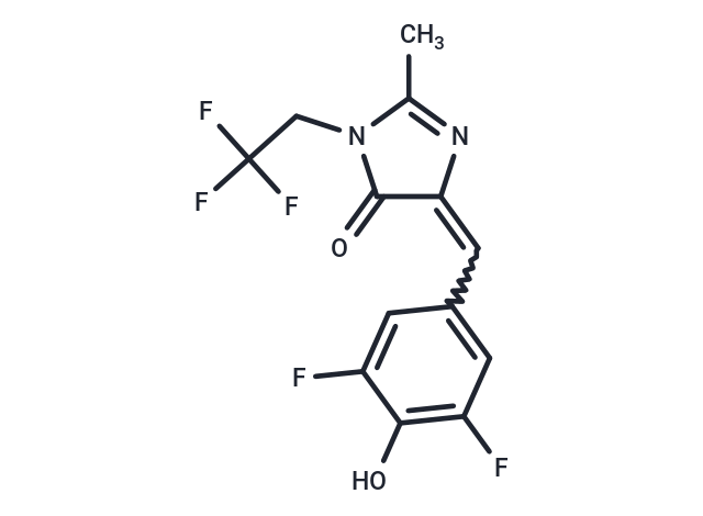
| Formula | C13H9F5N2O2 |
| Cas No. | 1539318-36-9 |
| Smiles | CC1=NC(=Cc2cc(F)c(O)c(F)c2)C(=O)N1CC(F)(F)F |
| Relative Density. | 1.49 g/cm3 (Predicted) |
| Storage | Keep away from direct sunlight,Keep away from moisture Powder: -20°C for 3 years | In solvent: -80°C for 1 year Shipping with blue ice/Shipping at ambient temperature. | |||||||||||||||||||||||||||||||||||
| Solubility Information | DMSO: 101 mg/mL (315.42 mM), Sonication is recommended. | |||||||||||||||||||||||||||||||||||
Solution Preparation Table | ||||||||||||||||||||||||||||||||||||
DMSO
Note : The dilution table applies only to solid products. For liquid products, please calculate the stock solution based on the stated concentration and/or density. | ||||||||||||||||||||||||||||||||||||
 For example, if the intended dosage is 10 mg/kg for animals weighing 20 g , with a dosing volume of 100 μL per animal, and a total of 10 animals are to be administered, using a formulation of
  10% DMSO+ 40% PEG300+ 5% Tween 80+ 45% Saline/PBS/ddH2O , the resulting working solution concentration would be 2 mg/mL.
Dissolve 2 mg of the compound in 100 μL DMSO to obtain a stock solution at a concentration of 20 mg/mL . If the required concentration exceeds the compound's known solubility, please contact us for technical support before proceeding.
1) Add 100 μL of the DMSO stock solution to 400 µL PEG300 and mix thoroughly until the solution becomes clear.
2) Add 50 µL Tween 80 and mix well until fully clarified.
3) Add 450 µL Saline,PBS or ddH2O and mix thoroughly until a homogeneous solution is obtained.
| Size | Quantity | Unit Price | Amount | Operation |
|---|
 Hello! How can I help you today?
 Copyright © 2015-2026 TargetMol Chemicals Inc. All Rights Reserved.
