
Shopping Cart
Remove All Your shopping cart is currently empty

Synonyms:
| Pack Size | Price | USA Stock | Global Stock | Quantity |
|---|---|---|---|---|
| 25 mg | $40 | - | In Stock | |
| 50 mg | $60 | Inquiry | Inquiry | |
| 1 mL x 10 mM (in DMSO) | $39 | In Stock | In Stock |
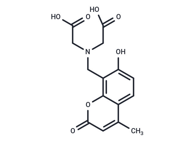
| Description | Calcein Blue is a coumarin-based fluorescent dye containing an iminodiacetic acid moiety that typically does not readily cross cell membranes. It can be used as a fluorescent labeling agent or as a metal ion fluorescent indicator, and is widely used in metal ion detection, cell imaging, and bioanalytical research. |
| In vitro | Calcein Blue (CB) forms a Calcein Blue–Fe²⁺ complex with Fe²⁺, and its fluorescence signal is significantly reduced due to the quenching effect of Fe²⁺. When dopamine (DA) is added to this system, it competes with Fe²⁺ for coordination, forming a more stable DA–Fe²⁺ complex, thereby releasing free CB and restoring its fluorescence. Under excitation at 340 nm and emission at 440 nm, the degree of fluorescence recovery shows a good linear correlation with dopamine concentration, making it suitable for the quantitative detection of dopamine. Among the catecholamines and related compounds studied, the Calcein Blue–Fe²⁺ system exhibits good selective recognition of dopamine [2]. |
| Molecular Weight | 321.28 |
| Formula | C15H15NO7 |
| Cas No. | 54375-47-2 |
| Smiles | Cc1cc(=O)oc2c(CN(CC(O)=O)CC(O)=O)c(O)ccc12 |
| Relative Density. | 1.512 g/cm3 |
| Storage | Keep away from direct sunlight Powder: -20°C for 3 years | In solvent: -80°C for 1 year Shipping with blue ice/Shipping at ambient temperature. | ||||||||||||||||||||
| Solubility Information | DMSO: 6 mg/mL (18.68 mM), Sonication and heating are recommended. | ||||||||||||||||||||
Solution Preparation Table | |||||||||||||||||||||
DMSO
Note : The dilution table applies only to solid products. For liquid products, please calculate the stock solution based on the stated concentration and/or density. | |||||||||||||||||||||
 For example, if the intended dosage is 10 mg/kg for animals weighing 20 g , with a dosing volume of 100 μL per animal, and a total of 10 animals are to be administered, using a formulation of
  10% DMSO+ 40% PEG300+ 5% Tween 80+ 45% Saline/PBS/ddH2O , the resulting working solution concentration would be 2 mg/mL.
Dissolve 2 mg of the compound in 100 μL DMSO to obtain a stock solution at a concentration of 20 mg/mL . If the required concentration exceeds the compound's known solubility, please contact us for technical support before proceeding.
1) Add 100 μL of the DMSO stock solution to 400 µL PEG300 and mix thoroughly until the solution becomes clear.
2) Add 50 µL Tween 80 and mix well until fully clarified.
3) Add 450 µL Saline,PBS or ddH2O and mix thoroughly until a homogeneous solution is obtained.
| Size | Quantity | Unit Price | Amount | Operation |
|---|
 Hello! How can I help you today?
 Copyright © 2015-2026 TargetMol Chemicals Inc. All Rights Reserved.
