
Shopping Cart
Remove All Your shopping cart is currently empty

Synonyms:

| Pack Size | Price | USA Stock | Global Stock | Quantity |
|---|---|---|---|---|
| 1 mg | $30 | In Stock | In Stock | |
| 5 mg | $74 | In Stock | In Stock | |
| 10 mg | $108 | In Stock | In Stock | |
| 25 mg | $178 | In Stock | In Stock | |
| 50 mg | $268 | In Stock | In Stock | |
| 100 mg | $398 | In Stock | In Stock | |
| 200 mg | $592 | - | In Stock |
| Description | DiD perchlorate is a lipophilic cyanine dye that can be used in cells tracking. |
| In vivo | Two weeks post-injection of stained cells, we observed a singular, luminous DiD perchlorate (DiD)-positive cell situated 15 to 40 μm from the endosteum. Our findings indicate a gradual emergence of cell clusters exhibiting diminished dye intensity, which aligns with the distribution of DiD perchlorate labeling upon cell division[1]. |
| Cell Research | I. Cell tracking 1. Solution preparation: DiD is usually dissolved in a solvent such as dimethyl sulfoxide (DMSO) to prepare a stock solution with a concentration of 1 to 10 μM. 2. Labeling: DiD solution is added to cultured cells. After 10 to 30 minutes of incubation, the dye will be absorbed by the cells and integrated into the lipid bilayer. After incubation, wash away the unbound dye with PBS. 3. Imaging: Observe using a fluorescence microscope or flow cytometer. DiD emits in the far-red band (665-680 nm) and can be used with other fluorescent markers for multiple experiments. 4. Cell tracking: DiD-labeled cells can be tracked in vitro or in vivo, and researchers can observe cell migration, proliferation, or localization in tissues. II. Membrane labeling 1. Membrane labeling: DiD binds to the cell membrane and can label cell surface or membrane proteins to observe the structure and dynamic changes of the membrane. 2. Imaging: Observe the labeled cells with a fluorescence microscope and analyze the distribution and changes of membrane proteins. III. Flow cytometry 1. Cell preparation: Label the cells as described above. 2. Flow cytometry: Use a flow cytometer to analyze the fluorescence intensity of labeled cells. Since DiD emits in the far-red range, it can be used with other common fluorescent probes for multiple labeling analysis. IV. Studying lipid raft dynamics 1. Labeling lipid rafts: DiD is able to bind to lipid rafts and can be used to monitor their organization in various cellular processes, such as cell signaling, membrane fusion, and endocytosis. 2. Analysis: After labeling the cells, use a fluorescence microscope to observe changes in lipid rafts after stimulation. The above information is based on published literature. Experimental procedures should be appropriately modified to meet specific research demands. |
| Molecular Weight | 959.9 |
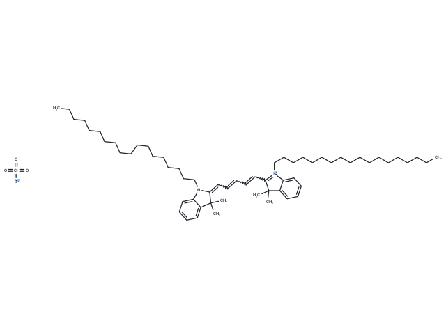
| Formula | C61H99ClN2O4 |
| Cas No. | 127274-91-3 |
| Smiles | [O-][Cl](=O)(=O)=O.CCCCCCCCCCCCCCCCCCN1C(=CC=CC=CC2=[N+](CCCCCCCCCCCCCCCCCC)c3ccccc3C2(C)C)C(C)(C)c2ccccc12 |
| Relative Density. | 1.31g/cm3 |
| Storage | Keep away from direct sunlight Powder: -20°C for 3 years Shipping with blue ice/Shipping at ambient temperature. | |||||||||||||||||||||||||||||||||||
| Solubility Information | DMSO: 50 mg/mL (52.09 mM), Sonication is recommended. Chloroform: 10 mg/mL (10.42 mM), Sonication is recommended. | |||||||||||||||||||||||||||||||||||
| In Vivo Formulation | 10% DMSO+40% PEG300+5% Tween-80+45% Saline: 1.67 mg/mL (1.74 mM), Sonication is recommended. Please add the solvents sequentially, clarifying the solution as much as possible before adding the next one. Dissolve by heating and/or sonication if necessary. Working solution is recommended to be prepared and used immediately. The formulation provided above is for reference purposes only. In vivo formulations may vary and should be modified based on specific experimental conditions. | |||||||||||||||||||||||||||||||||||
Solution Preparation Table | ||||||||||||||||||||||||||||||||||||
Chloroform/DMSO
DMSO
Note : The dilution table applies only to solid products. For liquid products, please calculate the stock solution based on the stated concentration and/or density. | ||||||||||||||||||||||||||||||||||||
 For example, if the intended dosage is 10 mg/kg for animals weighing 20 g , with a dosing volume of 100 μL per animal, and a total of 10 animals are to be administered, using a formulation of
  10% DMSO+ 40% PEG300+ 5% Tween 80+ 45% Saline/PBS/ddH2O , the resulting working solution concentration would be 2 mg/mL.
Dissolve 2 mg of the compound in 100 μL DMSO to obtain a stock solution at a concentration of 20 mg/mL . If the required concentration exceeds the compound's known solubility, please contact us for technical support before proceeding.
1) Add 100 μL of the DMSO stock solution to 400 µL PEG300 and mix thoroughly until the solution becomes clear.
2) Add 50 µL Tween 80 and mix well until fully clarified.
3) Add 450 µL Saline,PBS or ddH2O and mix thoroughly until a homogeneous solution is obtained.
| Size | Quantity | Unit Price | Amount | Operation |
|---|
 Hello! How can I help you today?
 Copyright © 2015-2026 TargetMol Chemicals Inc. All Rights Reserved.
