
Shopping Cart
Remove All Your shopping cart is currently empty

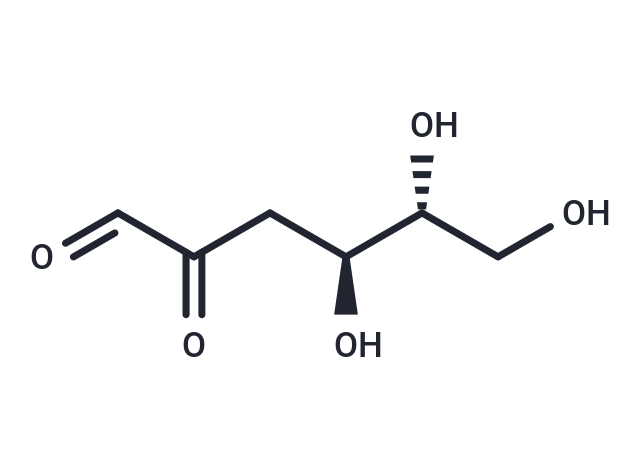
Synonyms: 3-Deoxy-D-glucosone, 2-keto-3-Deoxyglucose
| Pack Size | Price | USA Stock | Global Stock | Quantity |
|---|---|---|---|---|
| 1 mg | $156 | - | In Stock |
| Description | 3-Deoxyglucosone(3-Deoxy-D-glucosone) is synthesized by the intermediate pathway of the melad and polyol reactions.3-Deoxyglucosone reacts rapidly with protein amino groups to form advanced glycosylated end proteins (AGEs).3-Deoxyglucosone inactivates glutathione peroxidase and synergizes with low glucose to enhance GLP-1 secretion.3-Deoxyglucosone is used as a biomarker for diabetes mellitus.3-Deoxyglucosone has been shown to be an inhibitor of GLP-1 secretion in the presence of low glucose. 3-Deoxyglucosone inactivates glutathione oxidase, synergizes with low glucose to enhance GLP-1 secretion, and can be used as a biologic marker for diabetes. |
| In vitro | 3-Deoxyglucosone (80 ng/ml-1000 ng/ml; 1 hour; STC-1 cells) induces a 1.23-fold increase in GLP-1 secretion at 300 ng/ml and 1000 ng/ml, while 80 ng/ml shows no effect. At 300 ng/ml, 3DG significantly elevates intracellular Ca2+ levels, as measured by Fluo-3/AM (2.5 μM for 30 mins), but does not affect intracellular cAMP in a cAMP Elisa assay. Additionally, 300 ng/ml 3DG significantly enhances TAS1R2, TAS1R3, and TRPM5 protein expression under both glucose-free and high conditions.[1] |
| In vivo | 3-Deoxyglucosone (20 mg/kg; intragastric administration; single dose) induces a deterioration in glucose tolerance, as evidenced by an increased Area Under the Curve (AUC). Plasma glucagon levels do not show significant differences. The administration of 3-Deoxyglucosone leads to the development of impaired glucose regulation (IGR) accompanied by evident dysfunction of pancreatic islet cells in both Kunming mice and SD rats.[2] 3-deoxyglucosone (5-50 mg/kg; gastric gavage; once daily; 2 weeks; SD rats) exhibits significant increases in the upper small intestine (1.4-fold), lower small intestine (1.4-fold), ileum (1.4-fold), and colon (two-fold) compared to basal levels in their respective control groups. Additionally, there is a noteworthy decrease in the protein expressions of TAS1R2, TAS1R3, and TRPM5 observed in both the duodenum and colon.[3] |
| Synonyms | 3-Deoxy-D-glucosone, 2-keto-3-Deoxyglucose |
| Cell Research | Cell Line: STC-1 cells. Concentration: 300?ng/ml. Incubation Time: 1 hour [1] |
| Animal Research | Animal Model: SD rats. Dosage: 5, 20 and 50 mg/kg. Administration: oral administration; once daily; 2 weeks [3] |
| Molecular Weight | 162.14 |
| Formula | C6H10O5 |
| Cas No. | 4084-27-9 |
| Smiles | [C@H](CC(C=O)=O)([C@@H](CO)O)O |
| Relative Density. | 1.406g/cm3 |
| Storage | Store at low temperature Powder: -20°C for 3 years | In solvent: -80°C for 1 year Shipping with blue ice/Shipping at ambient temperature. |
 For example, if the intended dosage is 10 mg/kg for animals weighing 20 g , with a dosing volume of 100 μL per animal, and a total of 10 animals are to be administered, using a formulation of
  10% DMSO+ 40% PEG300+ 5% Tween 80+ 45% Saline/PBS/ddH2O , the resulting working solution concentration would be 2 mg/mL.
Dissolve 2 mg of the compound in 100 μL DMSO to obtain a stock solution at a concentration of 20 mg/mL . If the required concentration exceeds the compound's known solubility, please contact us for technical support before proceeding.
1) Add 100 μL of the DMSO stock solution to 400 µL PEG300 and mix thoroughly until the solution becomes clear.
2) Add 50 µL Tween 80 and mix well until fully clarified.
3) Add 450 µL Saline,PBS or ddH2O and mix thoroughly until a homogeneous solution is obtained.
| Size | Quantity | Unit Price | Amount | Operation |
|---|
 Hello! How can I help you today?
 Copyright © 2015-2026 TargetMol Chemicals Inc. All Rights Reserved.
