
Shopping Cart
Remove All Your shopping cart is currently empty

Synonyms: Kp76

| Pack Size | Price | USA Stock | Global Stock | Quantity |
|---|---|---|---|---|
| 1 mg | $48 | - | In Stock | |
| 5 mg | $112 | - | In Stock | |
| 10 mg | $163 | - | In Stock | |
| 25 mg | $248 | - | In Stock | |
| 50 mg | $370 | - | In Stock |
| Description | Kp7-6 is a Fas mimetic peptide that disrupts receptor complexes by simultaneously binding to Fas and FasL, thereby blocking downstream apoptotic signaling. Kp7-6 inhibits ERK1-2 phosphorylation, induces IκBα phosphorylation, and activates NF-κB. Kp7-6 suppresses the activation of caspase-8, caspase-3, and JNK, reduces human amylin-induced β-cell apoptosis, and decreases FasL-mediated lymphocyte cytotoxicity and apoptosis. In pancreatic neuroendocrine tumor models, Kp7-6 downregulates local FasL expression, increases CD8+Fas+ T-cell infiltration, and reduces tumor volume. Kp7-6 prevents concanavalin A-induced liver injury in mice and is applicable to research on type 2 diabetes, hepatitis, and pancreatic neuroendocrine tumors. |
| In vitro | Method: Isolated FVB/N mouse islets and RINm5F and CM cells were pretreated with Kp7-6 (0.5–5 mmol/L) for 1 h prior to exposure to hA (10 μmol/L or 40 μmol/L). Apoptosis was measured by cell death detection ELISA. In rescue experiments, Kp7-6 (5–10 mmol/L) was added 8 h after hA treatment. Circular dichroism spectroscopy and thioflavin-T binding assays were used to assess the effects of Kp7-6 on hA β-sheet and fibril formation. Western blotting was performed to detect caspase-8, caspase-3, and JNK protein expression levels. Result: Kp7-6 dose-dependently inhibited hA-induced apoptosis in islets, RINm5F, and CM cells. At 5 mmol/L, Kp7-6 completely suppressed apoptosis in cell lines but showed incomplete inhibition in isolated islets. Kp7-6 (5–10 mmol/L) added 8 h after hA treatment significantly rescued apoptosis, with similar effects in cell lines and islets. Kp7-6 (1:10 molar ratio to hA) inhibited hA β-sheet formation and prolonged the lag time of fibril formation to 5 h. Kp7-6 also suppressed the activation of downstream caspase-8, caspase-3, and JNK proteins[1]. Method: Human PNET cell lines BON-1 and QGP-1 were co-cultured with Jurkat T cells in the presence of Kp7-6. Apoptosis of Jurkat cells was assessed by flow cytometry (Annexin V/PI staining), and caspase-8/3 activities were measured using fluorometric activity assay kits. Result: Kp7-6 significantly reduced the proportion of apoptotic Jurkat T cells induced by ACSS2 overexpression and suppressed the elevation of caspase-8 and caspase-3 activities, indicating that Kp7-6 protects T cells from sFasL-mediated apoptosis by blocking Fas/FasL interactions[2]. Method: Human Jurkat T cells were co-treated with Kp7-6 and FasL-Flag for 24 h, and cell proliferation inhibition was measured by [³H]thymidine incorporation assay. In parallel, Jurkat cells were pretreated with Kp7-6 (1 mM) for 2 h followed by FasL (100 ng/mL) stimulation for indicated time periods, and phosphorylation levels of IκBα and ERK1/2 were detected by Western blotting. Result: Kp7-6 dose-dependently inhibited FasL-induced Jurkat cell killing, with >90% protection at high concentrations. Kp7-6 enhanced FasL-induced IκBα phosphorylation (at 5 min) while inhibiting ERK1/2 phosphorylation (at 5–30 min), thereby desensitizing cells to apoptotic signals through upregulation of NF-κB and suppression of the ERK pathway[3]. |
| In vivo | Method: In Rip1-Tag2 transgenic PNET spontaneous tumor mouse model (C57BL/6 background), Kp7-6 group received daily intraperitoneal injection of Kp7-6 (100 mg/kg, dissolved in 10% DMSO) from week 12, while the control group received an equivalent volume of vehicle. Multiplex immunofluorescence (mIF) staining was used to detect expression changes of ACSS2, FasL, Fas, and CD8 in tumor tissues, and tumor size and weight were measured. Result: In Kp7-6-treated mice, FasL expression in tumor tissues was significantly reduced, while infiltration of CD8⁺Fas⁺ T cells was markedly increased, with no change in ACSS2 expression. Tumor volume and weight were significantly lower than those in the control group, indicating that Kp7-6 restores CD8⁺ T cell infiltration and inhibits PNET tumor growth by antagonizing the Fas/FasL pathway[2]. Method: Eight-week-old C57BL/6 mice received intraperitoneal injection of Kp7-6 (3 mg/mouse) or control peptide Kp1-1 30 min before intravenous injection of Con A (0.5 mg/mouse). Blood samples were collected 12 h later, and serum alanine aminotransferase (ALT) and aspartate aminotransferase (AST) activities were measured using the Lippi-Guidi method to assess liver injury. Result: Compared with the Con A model group, serum ALT and AST levels in Kp7-6-pretreated mice were significantly reduced, while the control peptide Kp1-1 showed no protective effect, demonstrating that Kp7-6 effectively blocks Fas/FasL-mediated liver injury in vivo and provides significant protection against Con A-induced fulminant hepatitis[3]. |
| Synonyms | Kp76 |
| Molecular Weight | 1077.15 |
| Formula | C48H56N10O15S2 |
| Cas No. | 629628-53-1 |
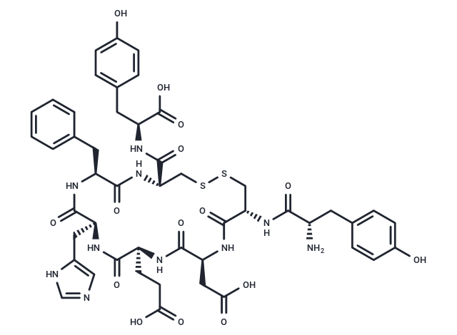
| Smiles | C([C@H]1C(=O)N[C@@H](CC2=CC=CC=C2)C(=O)N[C@H](C(N[C@@H](CC3=CC=C(O)C=C3)C(O)=O)=O)CSSC[C@H](NC([C@H](CC4=CC=C(O)C=C4)N)=O)C(=O)N[C@@H](CC(O)=O)C(=O)N[C@@H](CCC(O)=O)C(=O)N1)C5=CN=CN5 |
| Sequence | Tyr-Cys-Asp-Glu-His-Phe-Cys-Tyr (Disulfide bridge:Cys2-Cys7) |
| Sequence Short | YCDEHFCY (Disulfide bridge:Cys2-Cys7) |
| Storage | Keep away from moisture, Powder: -20°C for 3 years | In solvent: -80°C for 1 year Shipping with blue ice/Shipping at ambient temperature. | ||||||||||||||||||||||||||||||
| Solubility Information | DMSO: 80 mg/mL (74.27 mM), Sonication is recommended. | ||||||||||||||||||||||||||||||
Solution Preparation Table | |||||||||||||||||||||||||||||||
DMSO
Note : The dilution table applies only to solid products. For liquid products, please calculate the stock solution based on the stated concentration and/or density. | |||||||||||||||||||||||||||||||
 For example, if the intended dosage is 10 mg/kg for animals weighing 20 g , with a dosing volume of 100 μL per animal, and a total of 10 animals are to be administered, using a formulation of
  10% DMSO+ 40% PEG300+ 5% Tween 80+ 45% Saline/PBS/ddH2O , the resulting working solution concentration would be 2 mg/mL.
Dissolve 2 mg of the compound in 100 μL DMSO to obtain a stock solution at a concentration of 20 mg/mL . If the required concentration exceeds the compound's known solubility, please contact us for technical support before proceeding.
1) Add 100 μL of the DMSO stock solution to 400 µL PEG300 and mix thoroughly until the solution becomes clear.
2) Add 50 µL Tween 80 and mix well until fully clarified.
3) Add 450 µL Saline,PBS or ddH2O and mix thoroughly until a homogeneous solution is obtained.
| Size | Quantity | Unit Price | Amount | Operation |
|---|
 Hello! How can I help you today?
 Copyright © 2015-2026 TargetMol Chemicals Inc. All Rights Reserved.
