
Shopping Cart
Remove All Your shopping cart is currently empty

Synonyms: CBIC2

| Pack Size | Price | USA Stock | Global Stock | Quantity |
|---|---|---|---|---|
| 1 mg | $59 | In Stock | In Stock | |
| 2 mg | $81 | In Stock | In Stock | |
| 5 mg | $153 | In Stock | In Stock | |
| 10 mg | $252 | In Stock | In Stock | |
| 25 mg | $423 | In Stock | In Stock | |
| 50 mg | $628 | In Stock | In Stock | |
| 100 mg | $893 | - | In Stock |
| Description | JC-1 (CBIC2) is a fluorescent lipophilic cyanine dye. JC-1 is used to measure mitochondrial membrane potential. When the mitochondrial membrane potential is high, JC-1 aggregates into polymers within the matrix, emitting red fluorescence (λex=585 nm, λem=590 nm). When the mitochondrial membrane potential is low, JC-1 fails to aggregate within the mitochondrial matrix and exists as monomers, emitting green fluorescence (λex=510 nm, λem=527 nm). |
| Targets & IC50 | α-synuclein:2.6 μM (Kd) |
| In vitro | Methods: Primary cells from 12 AML patients (5 newly diagnosed, 7 relapsed) + 5 cell lines were treated with JC-1 (0.1 μM), Annexin V-FITC/PI assay kit, and cytarabine (10⁻⁵ M) + homoharringtonine (HHT, 1500 ng/mL) for apoptosis induction. After 48 hours of co-treatment, P-gp activity was compared between the two Methods via flow cytometry. Results: Following apoptosis induction, JC-1 red fluorescence remained significantly correlated with Rh 123 activity (P=0.01), whereas green fluorescence lost correlation (P=0.5), demonstrating red fluorescence as the superior marker for assessing P-gp activity.[1] |
| Synonyms | CBIC2 |
| Cell Research | Instructions I. Solution preparation 1. Stock solution preparation: JC-1 is dissolved in DMSO to prepare a stock solution. The concentration of the stock solution is usually 5 mg/ml. Please refer to the manufacturer's instructions for the specific concentration. 2. Working concentration: The working concentration of JC-1 is usually 1-20 µg/ml, but the optimal concentration may need to be optimized depending on the cell type and experimental conditions. 3. Dilution: Dilute the stock solution with cell culture medium or appropriate buffer before use. II. Staining cells 1. Cell preparation: Culture the cells to be tested under appropriate conditions (e.g., 37°C, 5% CO₂). 2. Incubation with JC-1: Add the diluted JC-1 working solution to the cells. Depending on the cell type and experimental conditions, incubate for 15-30 minutes. 3. Washing: After incubation, wash the cells 1-2 times with preheated cell culture medium or PBS to remove excess dye. 3. Fluorescence detection 1. Fluorescence microscopy: Use a fluorescence microscope equipped with appropriate filters to detect green (monomer JC-1) and red (aggregate JC-1) fluorescence. The excitation wavelength of green fluorescence is usually 485-490 nm, and that of red fluorescence is 525-530 nm, with emission peaks at 535 nm (green) and 590 nm (red), respectively. 2. Flow cytometry: Flow cytometry can also be used for quantitative analysis. Use appropriate filters (e.g., 525/40 nm for green fluorescence and 585/42 nm for red fluorescence) for detection. 3. Data interpretation: An increase in red fluorescence indicates a higher mitochondrial membrane potential, while an increase in green fluorescence indicates a loss or decrease in membrane potential. The above information is based on published literature. Experimental procedures should be appropriately modified to meet specific research demands. |
| Molecular Weight | 652.23 |
| Formula | C25H27Cl4IN4 |
| Cas No. | 3520-43-2 |
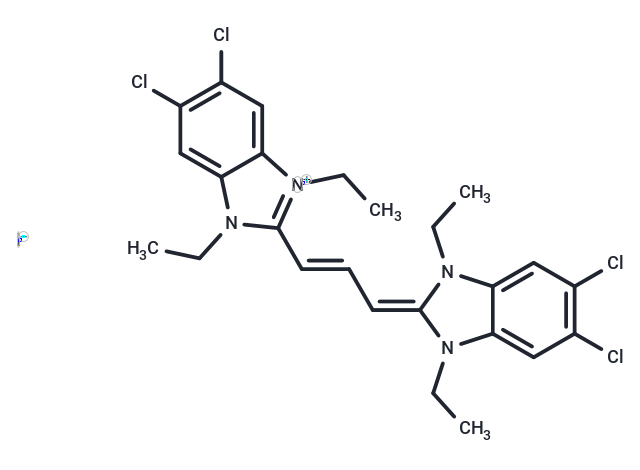
| Smiles | [I-].CCN1C(=C\C=C\c2n(CC)c3cc(Cl)c(Cl)cc3[n+]2CC)N(CC)c2cc(Cl)c(Cl)cc12 |
| Relative Density. | no data available |
| Storage | Keep away from direct sunlight,Store at low temperature,Keep away from moisture Powder: -20°C for 3 years | In solvent: -80°C for 1 year Shipping with blue ice/Shipping at ambient temperature. | |||||||||||||||||||||||||
| Solubility Information | DMSO: 14 mg/mL (21.46 mM), Sonication is recommended. | |||||||||||||||||||||||||
Solution Preparation Table | ||||||||||||||||||||||||||
DMSO
Note : The dilution table applies only to solid products. For liquid products, please calculate the stock solution based on the stated concentration and/or density. | ||||||||||||||||||||||||||
 For example, if the intended dosage is 10 mg/kg for animals weighing 20 g , with a dosing volume of 100 μL per animal, and a total of 10 animals are to be administered, using a formulation of
  10% DMSO+ 40% PEG300+ 5% Tween 80+ 45% Saline/PBS/ddH2O , the resulting working solution concentration would be 2 mg/mL.
Dissolve 2 mg of the compound in 100 μL DMSO to obtain a stock solution at a concentration of 20 mg/mL . If the required concentration exceeds the compound's known solubility, please contact us for technical support before proceeding.
1) Add 100 μL of the DMSO stock solution to 400 µL PEG300 and mix thoroughly until the solution becomes clear.
2) Add 50 µL Tween 80 and mix well until fully clarified.
3) Add 450 µL Saline,PBS or ddH2O and mix thoroughly until a homogeneous solution is obtained.
| Size | Quantity | Unit Price | Amount | Operation |
|---|
 Hello! How can I help you today?
 Copyright © 2015-2026 TargetMol Chemicals Inc. All Rights Reserved.
