
Shopping Cart
Remove All Your shopping cart is currently empty


Synonyms: DiIC18(3)
| Pack Size | Price | USA Stock | Global Stock | Quantity |
|---|---|---|---|---|
| 10 mg | $29 | In Stock | In Stock | |
| 1 mL x 10 mM (in DMSO) | $30 | In Stock | In Stock |
| Description | DiI (DiIC18(3)) is a lipophilic membrane dye commonly used as a long-term tracer for neurons and other cells. DiI fluoresces very weakly before it enters the cell membrane, and only after it enters the cell membrane can it be excited to emit a strong orange-red fluorescence (λex=549 nm, λem=565 nm). |
| In vitro | METHODS: DiI staining: 1. Prepare 1-5 mM of DiI dye stock solution in DMSO or EtOH. Protect from light and store in portions at -20°C. 2. 2. For use, dilute the stock solution with a physiological buffer (e.g., serum-free medium, HBSS, or PBS) to obtain a working solution of 1-5 µM DiI dye. The optimal working concentration must be determined empirically. 3. Suspend cells for staining: Suspend cells at a density of 1×10^6/mL in the dye working solution. Incubate at 37°C for 2-20 min. centrifuge labeled cells at 1000-1500 rpm for 5 min. remove supernatant and resuspend cells in pre-warmed growth medium. Repeat the washing twice. 4. Staining of adherent cells: Cultivate adherent cells on sterile coverslips. Remove the coverslip from the growth medium and gently drain off the excess medium. Pipette 100 μL of staining solution onto one corner of the coverslip and shake gently until all cells are covered. Incubate the coverslips at 37°C for 2-20 min. Drain the dye working solution and wash the coverslips two to three times with growth medium. For each wash, cover the cells with pre-warmed growth medium and incubate for 5-10 min, then drain the medium. 5. The stained cells can be examined by microscopy or flow cytometry. The above information is based on published literature. Experimental procedures should be appropriately modified to meet specific research demands. |
| Synonyms | DiIC18(3) |
| Cell Research | Instructions: I. Solution preparation 1. Mother solution preparation: Prepare DiL with DMSO to a concentration of 1-5 mM. 2. Working solution preparation: Dilute the mother solution to a suitable buffer such as serum-free culture medium, HBSS or PBS to prepare a working solution of 1 to 5 μM. II. Suspended cells 1. Centrifuge at 1000 g for 3-5 minutes at 4℃, discard the supernatant, and wash twice with PBS for 5 minutes each. The cell density is 1×10^6/mL. 2. Add 1 mL Di working solution and incubate at room temperature for 5-30 minutes. 3. Centrifuge at 400g for 3-4 minutes at 4℃ and discard the supernatant. 4. Wash twice with PBS for 5 minutes each. 5. Resuspend the cells with serum-free cell culture medium or PBS and observe with fluorescence microscopy or flow cytometry. III. Adherent cells 1. Culture adherent cells on sterile coverslips. 2. Remove the coverslip from the culture medium and aspirate the excess culture medium. 3. Add 100 μL of working solution, shake gently to completely cover the cells, and incubate at room temperature for 5-30 minutes. 4. Wash twice with culture medium, 5 minutes each time. Observe by fluorescence microscopy or flow cytometry. The above information is based on published literature. Experimental procedures should be appropriately modified to meet specific research demands. |
| Molecular Weight | 933.87 |
| Formula | C59H97ClN2O4 |
| Cas No. | 41085-99-8 |
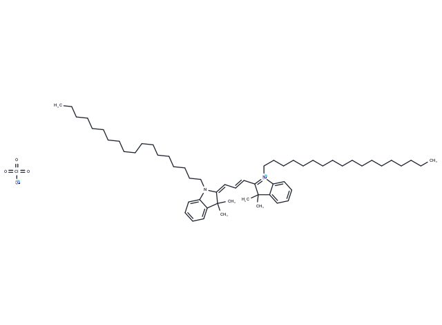
| Smiles | [O-][Cl](=O)(=O)=O.CCCCCCCCCCCCCCCCCCN1\C(=C\C=C\C2=[N+](CCCCCCCCCCCCCCCCCC)c3ccccc3C2(C)C)C(C)(C)c2ccccc12 |
| Relative Density. | no data available |
| Storage | Store at low temperature,Keep away from direct sunlight Powder: -20°C for 3 years | In solvent: -80°C for 1 year Shipping with blue ice/Shipping at ambient temperature. | ||||||||||||||||||||||||||||||
| Solubility Information | Chloroform: 10 mg/mL (10.71 mM), Sonication is recommended. DMSO: 39.6 mg/mL (42.4 mM), Sonication and heating are recommended. | ||||||||||||||||||||||||||||||
Solution Preparation Table | |||||||||||||||||||||||||||||||
Chloroform/DMSO
DMSO
Note : The dilution table applies only to solid products. For liquid products, please calculate the stock solution based on the stated concentration and/or density. | |||||||||||||||||||||||||||||||
 For example, if the intended dosage is 10 mg/kg for animals weighing 20 g , with a dosing volume of 100 μL per animal, and a total of 10 animals are to be administered, using a formulation of
  10% DMSO+ 40% PEG300+ 5% Tween 80+ 45% Saline/PBS/ddH2O , the resulting working solution concentration would be 2 mg/mL.
Dissolve 2 mg of the compound in 100 μL DMSO to obtain a stock solution at a concentration of 20 mg/mL . If the required concentration exceeds the compound's known solubility, please contact us for technical support before proceeding.
1) Add 100 μL of the DMSO stock solution to 400 µL PEG300 and mix thoroughly until the solution becomes clear.
2) Add 50 µL Tween 80 and mix well until fully clarified.
3) Add 450 µL Saline,PBS or ddH2O and mix thoroughly until a homogeneous solution is obtained.
| Size | Quantity | Unit Price | Amount | Operation |
|---|
 Hello! How can I help you today?
 Copyright © 2015-2026 TargetMol Chemicals Inc. All Rights Reserved.
