 Your shopping cart is currently empty


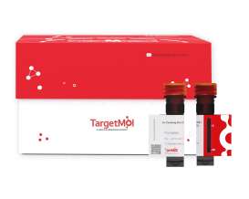
DiI Staining Solution
Once incorporated into the cell membrane, DiI can laterally diffuse, gradually labeling the entire membrane. Upon excitation, it emits bright orange-red fluorescence, with a maximum excitation wavelength of 549 nm and a maximum emission wavelength of 565 nm. The labeling of cell membranes by DiI is reversible; in the presence of unlabeled neighboring cells, DiI can be transferred between cells through membrane contact.
DiI is widely used as an anterograde or retrograde tracer for living or fixed neurons and other types of cells or tissues.
| Pack Size | Price | USA Warehouse | Global Warehouse | Quantity |
|---|---|---|---|---|
| 1 mL | $97 | In Stock | In Stock |
 Product Information
| DiI Staining Solution | Specifications |
|---|---|
| Ingredient | DiI |
| CAS | 41085-99-8 |
| Conc. | 5 mM |
| Solvent | DMSO |
 Features
1.Exhibits high specificity for cell membrane labeling: with short incubation time, it scarcely enters the cytoplasm, avoiding nuclear staining or interference from cellular organelles.
2.Suitable for long-term labeling for stable fluorescent signal.
3.Broad application: suitable for various sample types.
4.Simple operation & quick detection.
 Application
-
Cell membrane labeling & localization;
-
Analysis on cell membrane fluidity;
-
Analysis on cell division and morphology.
 Preparation of Working Solution
Dilute the DiI stock solution with an appropriate diluent (serum-free culture medium, PBS, or HBSS) to prepare the staining working solution (1-30 µM). The exact DiI working concentration should be adjusted according to the experimental conditions, with a typical concentration of 5-10 µM commonly used for cell membrane fluorescent labeling.
 Instructions
Staining of Suspension Live Cells
(1)Collect suspended cells in the logarithmic growth phase and centrifuge at 1,000 rpm, room temperature, for 5 min. Discard the supernatant. Resuspend in PBS, centrifuge again at 1,000 rpm, room temperature, for 5 min, and discard the supernatant.
(2)Resuspend cells in pre-warmed (37 ℃) culture medium and adjust cell density to 1×10⁶–5×10⁶ cells/mL.
(3)Add DiI working solution at 1/10 of the cell suspension volume, mix gently, and incubate at 37 ℃ in the dark for 5-20 min.
(4)Centrifuge at 1000 rpm for 5 min, discard the supernatant, and wash cells 2-3 times with pre-warmed (37 ℃) culture medium. Resuspend the cells.
(5)Analyze by flow cytometry, or drop the cell suspension onto a glass slide, cover with a coverslip, and observe under a fluorescence microscope. Excitation ≈ 549 nm, Emission ≈ 565 nm.
Staining of Adherent Live Cells
(1)Remove the culture medium and wash cells once with pre-warmed (37 ℃) culture medium.
(2)Add an appropriate amount of DiI working solution to cover all cells and incubate at 37 ℃ in the dark for 5-20 min.
(3)Remove the DiI solution and wash cells 2–3 times with pre-warmed (37 ℃) culture medium, 5 min each wash.
(4)Add pre-warmed (37 ℃) culture medium to cover the cells and observe under a fluorescence microscope. Excitation ≈ 549 nm, Emission ≈ 565 nm.
 Storage
Store at -20 ℃, protected from light for 12 months.
 Precautions
1.For staining live cells, it is recommended to use DiI at 37°C; for fixed cells or tissues, staining can be performed at room temperature. If fixation is required, 4% paraformaldehyde is generally recommended as the fixative.
2.During suspension cell staining, gently swirl the centrifuge tube every 10 minutes to prevent cells from settling and clumping, which may affect staining uniformity.
3.Since sample type and experimental conditions can affect staining efficiency, it is recommended to perform a pilot experiment to optimize the working solution concentration and staining time.
4.DiI is unstable in aqueous solutions; prolonged storage can lead to aggregation and decreased fluorescence efficiency. It is recommended to prepare the DiI working solution fresh before use.
5.Cells in the logarithmic growth phase have high viability and membrane fluidity. It is recommended to stain cells in the logarithmic phase and avoid using senescent or overly dense cells, as impaired membrane function can affect dye binding.
6.The product is for R&D use only, not for diagnostic procedures, food, drug, household or other uses.
7.Please wear a lab coat and disposable gloves.
 Instruction Manual
| Size | Quantity | Unit Price | Amount | Operation |
|---|

Copyright © 2015-2026 TargetMol Chemicals Inc. All Rights Reserved.


