
Shopping Cart
Remove All Your shopping cart is currently empty

Synonyms:
| Pack Size | Price | USA Stock | Global Stock | Quantity |
|---|---|---|---|---|
| 1 mg | $98 | - | In Stock | |
| 5 mg | $289 | - | In Stock | |
| 10 mg | $428 | - | In Stock | |
| 25 mg | $698 | - | In Stock | |
| 50 mg | $968 | - | In Stock |
| Description | Ginsenoside Rh3 is a natural product extracted from Ginseng C. A. Mey.Ginsenoside Rh3 has antifungal and antioxidant activities and induces Nrf2 activation in human retinal cells. |
| In vitro | Ginsenoside Rh3, at concentrations ranging from 0.01 to 10 μM, exhibits no cytotoxicity in tested models. In retinal pigment epithelium cells (RPEs), Ginsenoside Rh3 activates the Nrf2 pathway, as evidenced by dose-dependent increases in mRNA and protein levels of Nrf2-regulated genes, such as HO1, NQO1, and GCLC, following treatment. This enhancement in gene expression indicates a potentiation of cellular antioxidative mechanisms without altering Nrf2 mRNA levels, although Nrf2 protein levels significantly rise post-treatment with Ginsenoside Rh3 (3-10 μM). Additionally, the viability of SP 1-keratinocytes in response to Ginsenoside Rh3 has been evaluated using the EZ-Cytox assay, further delineating its safety and therapeutic potential. |
| In vivo | Ginsenoside Rh3 (5 mg/kg; intravitreal injection; 30 min pre-treatment) significantly attenuates light-induced decrease of both a- and b-wave amplitude. The electroretinography (ERG)'s a-wave decreases to 46.03±1.62% % of the control level after light exposure, which is back to 71.84±7.51% with Ginsenoside Rh3 administration. The b-wave is 40.19±3.34% of the control level by light exposure, and Rh3 intravitreal injection brings back to 80.01±2.37% of the control level.[1] |
| Cell Research | SP-1 keratinocytes are seeded in 96 well plates (2×10^4 cells/well). After 24 h, the media is replaced with media containing various concentrations of (A) SKRG, or (B) Ginsenoside Rh3 (0.01, 0.1, 1 and 10 μM). Control cells are treated with DMSO at a final concentration of 0.1%. After 24 h, the media containing the compounds or DMSO is replaced with media containing 10% EZ-Cytox. The cells are then incubated at 37°C for 1 h, and the absorbance is measured using a microplate reader at a wavelength of 450 nm. All assays are performed in triplicate [2]. |
| Animal Research | The BALB/c mice (male, 5-6 week old, 17-18 g weight) are used. The pupillary dilation is performed before exposure to 5000 lx of white fluorescent light. Thirty min before light exposure, Ginsenoside Rh3 (at 5 mg/kg body weight) is injected intravitreally to the right eye. ERG recording after light exposure is also reported early. The b-wave amplitude is measured from the trough of the a-wave to the peak of the b-wave, and the amplitude of the a-wave is measured from the initial baseline [1]. |
| Molecular Weight | 604.86 |
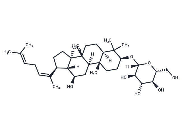
| Formula | C36H60O7 |
| Cas No. | 105558-26-7 |
| Smiles | [H][C@@]12[C@H](CC[C@@]1(C)[C@]1(C)CC[C@@]3([H])C(C)(C)[C@H](CC[C@]3(C)[C@@]1([H])C[C@H]2O)O[C@]1([H])O[C@H](CO)[C@@H](O)[C@H](O)[C@H]1O)C(\C)=C/CC=C(C)C |
| Relative Density. | 1.17 g/cm3 (Predicted) |
| Storage | Store at low temperature,Keep away from direct sunlight,Keep away from moisture Powder: -20°C for 3 years | In solvent: -80°C for 1 year Shipping with blue ice/Shipping at ambient temperature. | ||||||||||||||||||||||||||||||
| Solubility Information | Ethanol: 25 mg/mL (41.33 mM), Sonication is recommended. DMSO: 3.02 mg/mL (4.99 mM), Sonication and heating are recommended. | ||||||||||||||||||||||||||||||
Solution Preparation Table | |||||||||||||||||||||||||||||||
DMSO/Ethanol
Ethanol
Note : The dilution table applies only to solid products. For liquid products, please calculate the stock solution based on the stated concentration and/or density. | |||||||||||||||||||||||||||||||
 For example, if the intended dosage is 10 mg/kg for animals weighing 20 g , with a dosing volume of 100 μL per animal, and a total of 10 animals are to be administered, using a formulation of
  10% DMSO+ 40% PEG300+ 5% Tween 80+ 45% Saline/PBS/ddH2O , the resulting working solution concentration would be 2 mg/mL.
Dissolve 2 mg of the compound in 100 μL DMSO to obtain a stock solution at a concentration of 20 mg/mL . If the required concentration exceeds the compound's known solubility, please contact us for technical support before proceeding.
1) Add 100 μL of the DMSO stock solution to 400 µL PEG300 and mix thoroughly until the solution becomes clear.
2) Add 50 µL Tween 80 and mix well until fully clarified.
3) Add 450 µL Saline,PBS or ddH2O and mix thoroughly until a homogeneous solution is obtained.
| Size | Quantity | Unit Price | Amount | Operation |
|---|
 Hello! How can I help you today?
 Copyright © 2015-2026 TargetMol Chemicals Inc. All Rights Reserved.
