
Shopping Cart
Remove All Your shopping cart is currently empty

Synonyms:

| Pack Size | Price | USA Stock | Global Stock | Quantity |
|---|---|---|---|---|
| 1 mg | $35 | In Stock | In Stock | |
| 5 mg | $92 | In Stock | In Stock | |
| 10 mg | $139 | In Stock | In Stock | |
| 25 mg | $278 | In Stock | In Stock | |
| 50 mg | $438 | In Stock | In Stock | |
| 100 mg | $662 | In Stock | In Stock | |
| 200 mg | $926 | - | In Stock | |
| 1 mL x 10 mM (in DMSO) | $93 | In Stock | In Stock |
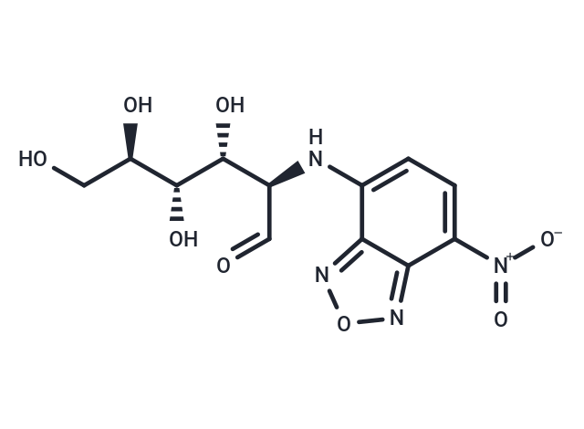
| Description | 2-NBDG is a fluorescent indicator for direct glucose uptake measurement. It is an indicator of cell viability. |
| In vitro | METHODS: Flow cytometry was used to detect glucose uptake: 1, Cells were seeded at 1*104/well in 96-well plates, and the experiment was performed within 24-48 h. The cells were incubated at 37 ℃ for 10-180 min. 2, Remove the cell culture medium, add fresh medium containing 2-NBDG (5-40 µM), and incubate for 10-180 min at 37 ℃. 3. Remove the medium and wash twice with pre-cooled PBS to stop the 2-NBDG uptake reaction. Resuspend in fresh medium and perform flow cytometry within 30 min. [1] The above information is based on published literature. Experimental procedures should be appropriately modified to meet specific research demands. |
| In vivo | METHODS: Glucose uptake by circulating breast cancer cells was detected by fluorescent microscopy: 1. A mouse blood sample (100 µL/mouse) was collected by puncturing the mouse saphenous vein. 2. Incubate the blood sample containing circulating breast cancer cells with 2-NBDG (5 µg/100 µL blood) for 30 min at 37℃ in a dark incubator. 3. Add the magnetic bead suspension (1µL 1%) to 100 µL of blood sample and incubate for 30 min at 4°C with gentle shaking to promote binding of the magnetic beads to the circulating breast cancer cells. 4. Separate the circulating breast cancer cells from the blood using a magnetic separation rack, wash with PBS 3 times, resuspend in 100 µL PBS and transfer to a 96-well cell plate. 5. 2-NBDG uptake by circulating breast cancer cells was examined under a fluorescence microscope equipped with a 488 nm filter. Large cells with a fluorescent signal derived from fluorescent 2-NBDG uptake by cells were counted as hypermetabolic circulating breast cancer cells, and small-sized normal mouse blood cells (lymphocytes and RBCs) showed no or little 2-NBDG fluorescent signal. [2] |
| Cell Research | Flow cytometry to detect glucose uptake Operation steps: a. Cells were seeded in 96-well plates at 1*104/well and the experiment was performed within 24-48 h; b. Cell culture medium was removed, fresh culture medium containing 2-NBDG (5-40 µM) was added, and incubated at 37 ℃ for 10-180 min; c. Culture medium was removed, and the cells were washed twice with pre-cooled PBS to stop the 2-NBDG uptake reaction. Resuspended in fresh culture medium, flow cytometry was performed within 30 min. The above information is based on published literature. Experimental procedures should be appropriately modified to meet specific research demands. |
| Molecular Weight | 342.26 |
| Formula | C12H14N4O8 |
| Cas No. | 186689-07-6 |
| Smiles | OC[C@@H](O)[C@@H](O)[C@H](O)[C@H](C=O)NC1=CC=C([N+]([O-])=O)C2=NON=C12 |
| Relative Density. | 1.750 g/cm3 (Predicted) |
| Storage | Store at low temperature Powder: -20°C for 3 years | In solvent: -80°C for 1 year Shipping with blue ice/Shipping at ambient temperature. | ||||||||||||||||||||||||||||||||||||||||
| Solubility Information | H2O: 5 mg/mL (14.61 mM), Sonication and heating to 60℃ are recommended. DMSO: 52.5 mg/mL (153.39 mM) | ||||||||||||||||||||||||||||||||||||||||
Solution Preparation Table | |||||||||||||||||||||||||||||||||||||||||
H2O/DMSO
DMSO
Note : The dilution table applies only to solid products. For liquid products, please calculate the stock solution based on the stated concentration and/or density. | |||||||||||||||||||||||||||||||||||||||||
 For example, if the intended dosage is 10 mg/kg for animals weighing 20 g , with a dosing volume of 100 μL per animal, and a total of 10 animals are to be administered, using a formulation of
  10% DMSO+ 40% PEG300+ 5% Tween 80+ 45% Saline/PBS/ddH2O , the resulting working solution concentration would be 2 mg/mL.
Dissolve 2 mg of the compound in 100 μL DMSO to obtain a stock solution at a concentration of 20 mg/mL . If the required concentration exceeds the compound's known solubility, please contact us for technical support before proceeding.
1) Add 100 μL of the DMSO stock solution to 400 µL PEG300 and mix thoroughly until the solution becomes clear.
2) Add 50 µL Tween 80 and mix well until fully clarified.
3) Add 450 µL Saline,PBS or ddH2O and mix thoroughly until a homogeneous solution is obtained.
| Size | Quantity | Unit Price | Amount | Operation |
|---|
 Hello! How can I help you today?
 Copyright © 2015-2026 TargetMol Chemicals Inc. All Rights Reserved.
