
Shopping Cart
Remove All Your shopping cart is currently empty

Synonyms:
| Pack Size | Price | USA Stock | Global Stock | Quantity |
|---|---|---|---|---|
| 1 mg | $30 | - | In Stock | |
| 5 mg | $58 | - | In Stock | |
| 10 mg | $84 | - | In Stock | |
| 25 mg | $138 | - | In Stock |
| Description | PDGFRA/CAIX/XII-IN-1 is a multi-target inhibitor targeting PDGFRA, CA IX and CA XII, with an IC50 of 20 nM against PDGFRA and Ki values of 93.3 nM and 80.0 nM against CA IX and CA XII respectively. It targets and binds to the ATP-binding pocket of PDGFRA, thereby blocking the activation of downstream signaling pathways such as STAT3, AKT and ERK1/2. It induces G0/G1 phase cell cycle arrest and initiates endogenous apoptotic procedures, manifested as cleavage and activation of PARP-1, caspase-9 and caspase-3, upregulation of caspase 3/7 activity and inhibition of Mcl-1 protein expression. It exerts significant anti-proliferative effects in eosinophilic leukemia cells and can serve as a candidate tool compound for mechanism and efficacy research of leukemia. |
| Targets & IC50 | CA IX:93.3 nM (Ki), PDGFRα (human):20 nM, CA XII:80.0 nM (Ki) |
| In vitro | Methods: The inhibitory activity of PDGFRA/CAIX/XII-IN-1 against recombinant PDGFRA protein and CA family isoforms was determined. Its targeting selectivity toward different RTK subfamilies was further explored. A variety of leukemia cell lines were treated with gradient concentrations for different durations, and the effects on cell proliferation, signaling pathways, apoptosis, and cell cycle progression were detected. Results: 1.PDGFRA/CAIX/XII-IN-1 potently inhibited recombinant PDGFRA protein with an IC₅₀ of 20 nM. At 1 μM, it exerted potent targeted inhibition against PDGFRα and closely related class III RTKs. 2.PDGFRA/CAIX/XII-IN-1 moderately inhibited recombinant human cancer-associated CA IX (Ki = 93.3 nM) and CA XII (Ki = 80.0 nM), while showing only weak inhibitory activity against normal CA I and CA II isoforms. 3.After 72 h treatment, PDGFRA/CAIX/XII-IN-1 strongly suppressed the proliferation of FIP1L1‑PDGFRA-driven EOL-1 cells (GI₅₀ = 2 nM). It displayed prominent cytotoxicity toward MV4-11 cells (GI₅₀ = 0.263 μM), whereas weaker cytotoxicity was observed in Kasumi-1 (GI₅₀ = 2.081 μM) and RS4-11 cells (GI₅₀ = 4.599 μM). 4.Treatment with 0–2500 nM for 1 h dose-dependently inhibited FIP1L1‑PDGFRA phosphorylation and downstream STAT3, AKT, and ERK1/2 signaling in EOL-1 cells; 100 nM treatment for 1 h achieved nearly complete PDGFRA inhibition. 5.Treatment with 0–2500 nM for 24 h induced EOL-1 cell apoptosis by activating the intrinsic apoptotic pathway. The pro-apoptotic effect was observed starting at 0.8 nM, with robust pathway activation at subnanomolar concentrations, while no pro-apoptotic effect was found in THP-1 cells. 6.Treatment with 0.16–100 nM for 24 h dose-dependently induced G0/G1 cell cycle arrest in EOL-1 cells, with a significant effect already evident at 0.8 nM. Concentrations of 4 nM and above induced apoptotic cell death accompanied by an increased sub-G1 population [1]. |
| Molecular Weight | 507.56 |
| Formula | C25H25N5O5S |
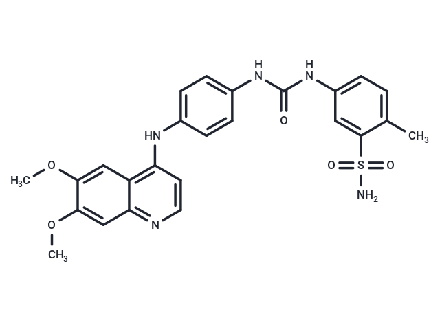
| Smiles | COC1=CC2=C(NC3=CC=C(NC(NC4=CC=C(C)C(S(N)(=O)=O)=C4)=O)C=C3)C=CN=C2C=C1OC |
| Relative Density. | no data available |
| Storage | Powder: -20°C for 3 years | In solvent: -80°C for 1 year Shipping with blue ice/Shipping at ambient temperature. |
 For example, if the intended dosage is 10 mg/kg for animals weighing 20 g , with a dosing volume of 100 μL per animal, and a total of 10 animals are to be administered, using a formulation of
  10% DMSO+ 40% PEG300+ 5% Tween 80+ 45% Saline/PBS/ddH2O , the resulting working solution concentration would be 2 mg/mL.
Dissolve 2 mg of the compound in 100 μL DMSO to obtain a stock solution at a concentration of 20 mg/mL . If the required concentration exceeds the compound's known solubility, please contact us for technical support before proceeding.
1) Add 100 μL of the DMSO stock solution to 400 µL PEG300 and mix thoroughly until the solution becomes clear.
2) Add 50 µL Tween 80 and mix well until fully clarified.
3) Add 450 µL Saline,PBS or ddH2O and mix thoroughly until a homogeneous solution is obtained.
| Size | Quantity | Unit Price | Amount | Operation |
|---|
 Hello! How can I help you today?
 Copyright © 2015-2026 TargetMol Chemicals Inc. All Rights Reserved.
