
Shopping Cart
Remove All Your shopping cart is currently empty


Synonyms:
| Pack Size | Price | USA Stock | Global Stock | Quantity |
|---|---|---|---|---|
| 25 mg | $29 | In Stock | In Stock | |
| 50 mg | $49 | In Stock | In Stock |
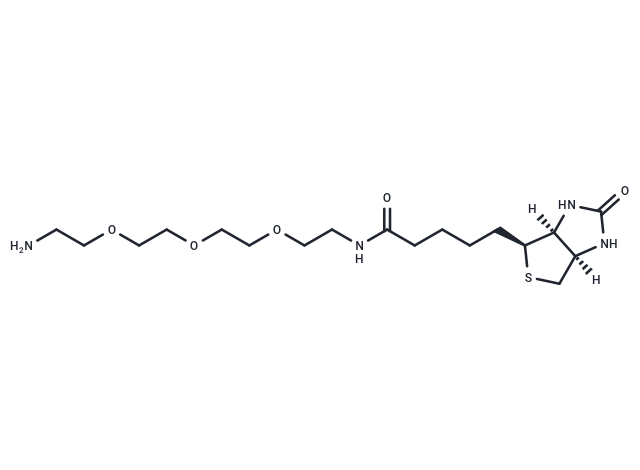
| Description | Amine-PEG3-Biotin can be used as a signal amplification label. |
| Cell Research | 1. Binding to biological molecules 1. Labeling reaction: Amine-PEG3-Biotin reacts with the target biological molecule, usually by amidation reaction to connect the dye molecule to the target molecule. Depending on the desired labeling effect, the reaction conditions, such as pH, reaction time and temperature, may need to be optimized. 2. Purification of labeled molecules: After the reaction is completed, unreacted biotin is usually removed by dialysis or chromatography. 3. Detection: Detection is performed using streptavidin-enzyme or streptavidin-fluorescent markers to amplify the signal for analysis. 2. Western Blot and immunoassay 1. Labeling antibodies: Enhance the detection sensitivity by labeling Amine-PEG3-Biotin with primary or secondary antibodies. 2. Detection: The labeled antibody is used for protein transfer and detection is performed using streptavidin-enzyme or streptavidin-fluorescent markers. 3. Signal amplification: The strong binding of biotin to streptavidin enables the signal to be amplified, improving the sensitivity of immunoassay. III. ELISA (Enzyme-Linked Immunosorbent Assay) 1. Coating: Coat the biotin-labeled target molecule onto the ELISA plate (such as a labeled antibody or antigen). 2. Detection: Detection is performed by adding streptavidin-enzyme conjugates and generating color or signal through substrate reaction. IV. Cell experiment 1. Labeling cells: Add Amine-PEG3-Biotin to the cell culture medium and incubate for a certain period of time (usually 30 minutes to 1 hour). 2. Washing: Wash away unbound dye with PBS. 3. Fluorescence microscopy: Use a fluorescence microscope or flow cytometer for detection to observe the staining pattern and dynamic process of the cells. The above information is based on published literature. Experimental procedures should be appropriately modified to meet specific research demands. |
| Molecular Weight | 418.551 |
| Formula | C18H34N4O5S |
| Cas No. | 359860-27-8 |
| Smiles | O=C(NCCOCCOCCOCCN)CCCC[C@@H]1SC[C@]([C@]1([H])N2)([H])NC2=O |
| Relative Density. | 1.166 g/cm3 (Predicted) |
| Storage | Keep away from direct sunlight Store at -20°C Shipping with blue ice/Shipping at ambient temperature. | |||||||||||||||||||||||||||||||||||
| Solubility Information | H2O: 95.00 mg/mL (226.97 mM), Sonication is recommended. DMSO: 245.00 mg/mL (585.35 mM), Sonication is recommended. | |||||||||||||||||||||||||||||||||||
| In Vivo Formulation | 10% DMSO+90% Saline: 10 mg/mL (23.89 mM), Solution. 10% DMSO+40% PEG300+5% Tween-80+45% Saline: 1.00 mg/mL (2.39 mM), Sonication is recommended. Please add the solvents sequentially, clarifying the solution as much as possible before adding the next one. Dissolve by heating and/or sonication if necessary. Working solution is recommended to be prepared and used immediately. The formulation provided above is for reference purposes only. In vivo formulations may vary and should be modified based on specific experimental conditions. | |||||||||||||||||||||||||||||||||||
Solution Preparation Table | ||||||||||||||||||||||||||||||||||||
H2O/DMSO
Note : The dilution table applies only to solid products. For liquid products, please calculate the stock solution based on the stated concentration and/or density. | ||||||||||||||||||||||||||||||||||||
 For example, if the intended dosage is 10 mg/kg for animals weighing 20 g , with a dosing volume of 100 μL per animal, and a total of 10 animals are to be administered, using a formulation of
  10% DMSO+ 40% PEG300+ 5% Tween 80+ 45% Saline/PBS/ddH2O , the resulting working solution concentration would be 2 mg/mL.
Dissolve 2 mg of the compound in 100 μL DMSO to obtain a stock solution at a concentration of 20 mg/mL . If the required concentration exceeds the compound's known solubility, please contact us for technical support before proceeding.
1) Add 100 μL of the DMSO stock solution to 400 µL PEG300 and mix thoroughly until the solution becomes clear.
2) Add 50 µL Tween 80 and mix well until fully clarified.
3) Add 450 µL Saline,PBS or ddH2O and mix thoroughly until a homogeneous solution is obtained.
| Size | Quantity | Unit Price | Amount | Operation |
|---|
 Hello! How can I help you today?
 Copyright © 2015-2026 TargetMol Chemicals Inc. All Rights Reserved.
