
Shopping Cart
Remove All Your shopping cart is currently empty

Synonyms:

| Pack Size | Price | USA Stock | Global Stock | Quantity |
|---|---|---|---|---|
| 5 mg | $57 | In Stock | In Stock | |
| 10 mg | $86 | In Stock | In Stock | |
| 25 mg | $174 | In Stock | In Stock | |
| 50 mg | $259 | In Stock | In Stock | |
| 100 mg | $388 | - | In Stock | |
| 1 mL x 10 mM (in DMSO) | $66 | In Stock | In Stock |
| Description | DiBAC4(3) is a voltage-sensitive fluorescent dye (λem=505 nm, λex=490 nm). |
| In vitro | Membrane hyperpolarization in HEKBKα, induced by 10 μM Evans blue (EB), is distinctly detected using DiBAC4(3), contrasting with the gradual alteration in membrane potential observed upon adding 3 mM tetraethylammonium chloride compared to microelectrode assessments. Hyperpolarization peak times are 2.3±0.9 s and 35.0±2.6 s, as measured with microelectrodes and DiBAC4(3), respectively. |
| Cell Research | Instructions I. Solution preparation 1. Stock solution: DiBAC4(3) can be dissolved in DMSO or other appropriate solvents to prepare a stock solution with a concentration of 1-10 mM. 2. Working solution: Dilute the stock solution to a working concentration, usually 1–5 µM, using an appropriate buffer or culture medium (e.g., PBS or RPMI) as needed for the experiment. II. Procedure 1. Cell staining: Seed the cells in a suitable culture dish and culture to the desired density. Add the DiBAC4(3) working solution to the culture medium containing the cells. 2. Incubate at 37°C for 15–30 minutes. The incubation time may vary depending on the experimental conditions. Avoid light during incubation to prevent dye degradation. 3. Washing: After incubation, wash the cells with fresh culture medium or PBS to remove unbound dye. 4. The cells can now be used for fluorescence detection. 5. Fluorescence detection: Excitation and emission wavelengths: DiBAC4(3) can be excited by light at a wavelength of 490 nm and emits light at approximately 505 nm. Fluorescence intensity is related to changes in membrane potential. Increased fluorescence intensity is generally associated with hyperpolarization, while decreased fluorescence intensity is associated with depolarization. Microscopy/flow cytometry: For fluorescence microscopy, a microscope equipped with appropriate filters is used to observe fluorescence at an emission wavelength of 505 nm; for flow cytometry, fluorescence can be measured in the 505 nm range to analyze changes in membrane potential of individual cells. 6. Application areas: 1) Membrane potential research: DiBAC4(3) is widely used to monitor changes in membrane potential of living cells, which is crucial for ion channel activity, drug screening, and cell excitability studies. 2) Cardiac and neural research: It is often used in electrophysiological studies of cardiac cells, neurons, and other excitable cells. 3) Toxicology and cell stress: DiBAC4(3) can also be used to evaluate the effects of toxic factors on cell membrane potential. Notes: 1. Photosensitivity: DiBAC4(3) is light sensitive and samples should be handled in the dark or in a low light environment. 2. Toxicity: Although generally safe at working concentrations, high concentrations may affect cell viability. Therefore, it is recommended to optimize the concentration for each cell type. The above information is based on published literature. Experimental procedures should be appropriately modified to meet specific research demands. |
| Molecular Weight | 516.63 |
| Formula | C27H40N4O6 |
| Cas No. | 70363-83-6 |
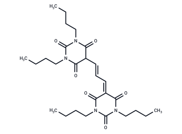
| Smiles | CCCCN1C(=O)C(\C=C\C=C2C(=O)N(CCCC)C(=O)N(CCCC)C2=O)C(=O)N(CCCC)C1=O |
| Relative Density. | 1.201 g/cm3 (Predicted) |
| Storage | Keep away from direct sunlight Powder: -20°C for 3 years | In solvent: -80°C for 1 year Shipping with blue ice/Shipping at ambient temperature. | |||||||||||||||||||||||||||||||||||
| Solubility Information | DMSO: 55 mg/mL (106.46 mM), Sonication is recommended. | |||||||||||||||||||||||||||||||||||
Solution Preparation Table | ||||||||||||||||||||||||||||||||||||
DMSO
Note : The dilution table applies only to solid products. For liquid products, please calculate the stock solution based on the stated concentration and/or density. | ||||||||||||||||||||||||||||||||||||
 For example, if the intended dosage is 10 mg/kg for animals weighing 20 g , with a dosing volume of 100 μL per animal, and a total of 10 animals are to be administered, using a formulation of
  10% DMSO+ 40% PEG300+ 5% Tween 80+ 45% Saline/PBS/ddH2O , the resulting working solution concentration would be 2 mg/mL.
Dissolve 2 mg of the compound in 100 μL DMSO to obtain a stock solution at a concentration of 20 mg/mL . If the required concentration exceeds the compound's known solubility, please contact us for technical support before proceeding.
1) Add 100 μL of the DMSO stock solution to 400 µL PEG300 and mix thoroughly until the solution becomes clear.
2) Add 50 µL Tween 80 and mix well until fully clarified.
3) Add 450 µL Saline,PBS or ddH2O and mix thoroughly until a homogeneous solution is obtained.
| Size | Quantity | Unit Price | Amount | Operation |
|---|
 Hello! How can I help you today?
 Copyright © 2015-2026 TargetMol Chemicals Inc. All Rights Reserved.
