
Shopping Cart
Remove All Your shopping cart is currently empty


Synonyms:

| Pack Size | Price | USA Stock | Global Stock | Quantity |
|---|---|---|---|---|
| 2 mg | $38 | In Stock | In Stock | |
| 5 mg | $61 | In Stock | In Stock | |
| 10 mg | $101 | In Stock | In Stock | |
| 25 mg | $215 | In Stock | In Stock | |
| 50 mg | $360 | In Stock | In Stock | |
| 100 mg | $535 | In Stock | In Stock | |
| 1 mL x 10 mM (in DMSO) | $65 | In Stock | In Stock |
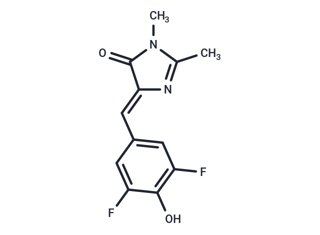
| Description | DFHBI, a small molecule resembling the chromophore of green fluorescent protein (GFP), is essentially nonfluorescent when unbound. The Spinach-DFHBI complex, however, exhibits bright fluorescence both in vitro and in living cells. |
| In vitro | Spinach and Spinach2 bind to DFHBI have fluorescence excitation maxima of 447 nm and peak fluorescence emission of 501 nm[1] and they are RNA aptamers that can be used for the genetic encoding of fluorescent RNA. Spinach is a 98-nt-long RNA aptamer that binds to and switches on the fluorescence of DFHBI. Both Spinach and DFHBI are essentially nonfluorescent when unbound, whereas the Spinach-DFHBI complex is brightly fluorescent both in vitro and in living cells. Spinach2 binds and activates the fluorescence of DFHBI, allowing the dynamic localizations of Spinach2-tagged RNAs to be imaged in live cells. The spectral properties of Spinach2 are limited by DFHBI, which produces fluorescence that is bluish-green and is not optimized for filters commonly used in fluorescence microscopes. DFHBI should be shielded from light. All stock solutions of DFHBI should be maintained in dark tubes or wrapped in foil. Plates containing cultures incubated with DFHBI should be kept in the dark by using a foil overwrap[2]. |
| Cell Research | I. RNA labeling and imaging 1. Prepare Spinach RNA: synthesize target RNA containing Spinach aptamer sequence by in vitro transcription or RNA synthesis method. 2. Dissolve DFHBI: dissolve DFHBI in DMSO or water to make a 1-10 mM stock solution, and dilute to 1-50 µM range when used. 3. Complex formation: mix Spinach RNA with DFHBI and incubate under appropriate buffer conditions (such as Tris-HCl, MgCl₂, etc.) to form a highly fluorescent complex. 3. Imaging: observe using a fluorescence microscope, with an excitation wavelength of 470-490 nm and an emission wavelength of 510-530 nm. II. Live cell experiment 1. Spinach RNA expression: transfect cells with a plasmid containing Spinach sequence, or express target RNA through an in vivo RNA synthesis system. 2. Add DFHBI: add DFHBI (final concentration is usually 10-50 µM) to the cell culture medium and incubate for 10-30 minutes. 3. Fluorescence detection: Observe under a fluorescence microscope or fluorescence imager to monitor the localization and dynamics of RNA. 4 Fluorescence detection and quantification 1) Fluorescence measurement: Use a fluorescence spectrophotometer to quantify the fluorescence intensity of the Spinach-DFHBI complex to analyze the amount or dynamic changes of RNA. 2) Optimization conditions: Optimize the buffer, ion concentration (especially Mg²⁺ concentration) and pH in the experiment to obtain the best fluorescence signal. The above information is based on published literature. Experimental procedures should be appropriately modified to meet specific research demands. |
| Molecular Weight | 252.22 |
| Formula | C12H10F2N2O2 |
| Cas No. | 1241390-29-3 |
| Smiles | CN1C(C)=N\C(=C/c2cc(F)c(O)c(F)c2)C1=O |
| Relative Density. | no data available |
| Storage | Keep away from direct sunlight Powder: -20°C for 3 years | In solvent: -80°C for 1 year Shipping with blue ice/Shipping at ambient temperature. | |||||||||||||||||||||||||||||||||||
| Solubility Information | DMSO: 85 mg/mL (337.01 mM), Sonication is recommended. | |||||||||||||||||||||||||||||||||||
| In Vivo Formulation | 10% DMSO+40% PEG300+5% Tween-80+45% Saline: 1 mg/mL (3.96 mM), Sonication is recommended. Please add the solvents sequentially, clarifying the solution as much as possible before adding the next one. Dissolve by heating and/or sonication if necessary. Working solution is recommended to be prepared and used immediately. The formulation provided above is for reference purposes only. In vivo formulations may vary and should be modified based on specific experimental conditions. | |||||||||||||||||||||||||||||||||||
Solution Preparation Table | ||||||||||||||||||||||||||||||||||||
DMSO
Note : The dilution table applies only to solid products. For liquid products, please calculate the stock solution based on the stated concentration and/or density. | ||||||||||||||||||||||||||||||||||||
 For example, if the intended dosage is 10 mg/kg for animals weighing 20 g , with a dosing volume of 100 μL per animal, and a total of 10 animals are to be administered, using a formulation of
  10% DMSO+ 40% PEG300+ 5% Tween 80+ 45% Saline/PBS/ddH2O , the resulting working solution concentration would be 2 mg/mL.
Dissolve 2 mg of the compound in 100 μL DMSO to obtain a stock solution at a concentration of 20 mg/mL . If the required concentration exceeds the compound's known solubility, please contact us for technical support before proceeding.
1) Add 100 μL of the DMSO stock solution to 400 µL PEG300 and mix thoroughly until the solution becomes clear.
2) Add 50 µL Tween 80 and mix well until fully clarified.
3) Add 450 µL Saline,PBS or ddH2O and mix thoroughly until a homogeneous solution is obtained.
| Size | Quantity | Unit Price | Amount | Operation |
|---|
 Hello! How can I help you today?
 Copyright © 2015-2026 TargetMol Chemicals Inc. All Rights Reserved.
