
Shopping Cart
Remove All Your shopping cart is currently empty

Synonyms:

| Pack Size | Price | USA Stock | Global Stock | Quantity |
|---|---|---|---|---|
| 1 mg | $58 | - | In Stock | |
| 5 mg | $177 | - | In Stock | |
| 10 mg | $283 | - | In Stock | |
| 25 mg | $478 | - | In Stock | |
| 50 mg | $698 | - | In Stock |
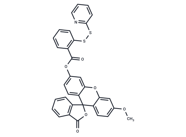
| Description | WSP-1 is a selective 2-pyridyl disulfide fluorescent probe for hydrogen sulfide detection and bioimaging. |
| Cell Research | 1. Hydrogen sulfide detection Experimental steps: 1. Prepare WSP-1 solution: WSP-1 is usually dissolved in an appropriate solvent (such as DMSO) and then mixed with cell culture or biological samples. Its concentration is usually between 1-5 μM (can be adjusted according to experimental requirements). 2. Incubate the cell/tissue sample: Add WSP-1 solution to live cells or tissues, usually 30-60 minutes, allowing the probe to enter the cells and react with hydrogen sulfide. 3. Fluorescence imaging: Use a fluorescence microscope or imaging system to monitor the fluorescence signal. WSP-1 emits fluorescence at a specific wavelength in the presence of hydrogen sulfide, usually within the green fluorescence range. 4. Data analysis: According to the fluorescence intensity, the concentration of hydrogen sulfide in cells or tissues can be quantitatively analyzed. 2. Application in cell and animal models Experimental steps: 1. Preparation of cell/animal model: Prepare appropriate cell lines or animal models, inject or add WSP-1 probes through cell culture. 2. Monitoring and imaging: Use a fluorescence microscope or multiphoton microscope to capture fluorescence images at appropriate time points to analyze the dynamic changes of hydrogen sulfide. 3. Results analysis: The concentration of hydrogen sulfide and its relationship with pathological status were evaluated through fluorescence images and quantitative analysis. 3. Research on hydrogen sulfide: Experimental steps: 1. Drug intervention or mutation model: Change hydrogen sulfide levels in cells or animals by adding synthesizers or inhibitors of hydrogen sulfide (such as inhibitors of certain hydrogen sulfide-generating enzymes). 2. Fluorescence monitoring: Monitor the changes in the fluorescence signal generated by WSP-1 to evaluate the concentration changes of hydrogen sulfide under different experimental conditions. 3. Results analysis: Fluorescence signals are used to quantitatively analyze the relationship between hydrogen sulfide and related physiological or pathological events (such as heart disease, neurodegenerative diseases, etc.). Notes: 1. Solubility and stability: WSP-1 has good solubility, but when used, it should avoid direct contact with strong reducing agents or high concentrations of hydrogen sulfide to avoid affecting its performance. 2. Background interference: Background fluorescence in certain cells or tissues may interfere with measurements. Imaging conditions should be optimized and controlled experiments should be conducted when used. 3. Cell permeability: WSP-1 has a high permeability and can directly enter the cells and react with endogenous hydrogen sulfide, but for some cell types, it may be necessary to optimize experimental conditions. The above information is based on published literature. Experimental procedures should be appropriately modified to meet specific research demands. |
| Molecular Weight | 591.65 |
| Formula | C33H21NO6S2 |
| Cas No. | 1352750-34-5 |
| Smiles | O=C(OC1=CC=C2C(OC3=CC(OC)=CC=C3C42OC(=O)C=5C=CC=CC54)=C1)C=6C=CC=CC6SSC7=NC=CC=C7 |
| Storage | Keep away from direct sunlight Powder: -20°C for 3 years | In solvent: -80°C for 1 year Shipping with blue ice/Shipping at ambient temperature. | ||||||||||||||||||||||||||||||
| Solubility Information | DMF: 20 mg/mL (33.8 mM), Sonication is recommended. DMSO: 8.53 mg/mL (14.42 mM), Sonication is recommended. | ||||||||||||||||||||||||||||||
Solution Preparation Table | |||||||||||||||||||||||||||||||
DMSO/DMF
DMF
Note : The dilution table applies only to solid products. For liquid products, please calculate the stock solution based on the stated concentration and/or density. | |||||||||||||||||||||||||||||||
 For example, if the intended dosage is 10 mg/kg for animals weighing 20 g , with a dosing volume of 100 μL per animal, and a total of 10 animals are to be administered, using a formulation of
  10% DMSO+ 40% PEG300+ 5% Tween 80+ 45% Saline/PBS/ddH2O , the resulting working solution concentration would be 2 mg/mL.
Dissolve 2 mg of the compound in 100 μL DMSO to obtain a stock solution at a concentration of 20 mg/mL . If the required concentration exceeds the compound's known solubility, please contact us for technical support before proceeding.
1) Add 100 μL of the DMSO stock solution to 400 µL PEG300 and mix thoroughly until the solution becomes clear.
2) Add 50 µL Tween 80 and mix well until fully clarified.
3) Add 450 µL Saline,PBS or ddH2O and mix thoroughly until a homogeneous solution is obtained.
| Size | Quantity | Unit Price | Amount | Operation |
|---|
 Hello! How can I help you today?
 Copyright © 2015-2026 TargetMol Chemicals Inc. All Rights Reserved.
