
Shopping Cart
Remove All Your shopping cart is currently empty

Synonyms:
| Pack Size | Price | USA Stock | Global Stock | Quantity |
|---|---|---|---|---|
| 5 mg | $30 | - | In Stock | |
| 10 mg | $39 | In Stock | In Stock | |
| 25 mg | $67 | - | In Stock | |
| 1 mL x 10 mM (in DMSO) | $50 | - | In Stock |
| Description | Rhodamine 800 is a near-infrared fluorescent dye. |
| In vitro | The fluorescence intensity of Rhodamine 800 varies markedly with the solvent and decreases to less than one-fifth when the solvent is changed from 2-propanone to water without a significant change of the emission peak. |
| Cell Research | Instructions I. Reagent preparation 1. Stock solution preparation: Dissolve it in an appropriate solvent, such as dimethyl sulfoxide (DMSO) or ethanol, to prepare a stock solution. The concentration of the stock solution is usually 0.1 mM to 1 mM, and the specific concentration depends on the experimental requirements. 2. Working solution preparation: When used in vitro or in vivo, the stock solution is usually diluted with PBS (phosphate buffered saline) or other suitable buffer to the required working concentration, usually ranging from 1 µM to 100 µM, and the specific concentration is adjusted according to the experimental design. II. Operation steps 1. Sample preparation: 1) Cell labeling: Rhodamine 800 can be used to label cells, tissues, or proteins. If labeling cells, the dye should be incubated with the cells for a period of time (usually 30 minutes to 1 hour), and then washed to remove excess dye. 2) In vivo imaging: In in vivo imaging experiments, Rhodamine 800 can be injected intravenously or applied topically. The dye will accumulate in specific tissues or organs, and fluorescence detection is performed using a near-infrared imaging system. 2. Labeling and imaging: 1) Cell culture: Incubate cells with Rhodamine 800 solution for typically 30-60 minutes, then wash cells with PBS to remove unbound dye. Analysis can then be performed using a near-infrared fluorescence microscope or flow cytometer. 2) Tissue and in vivo imaging: For tissue or in vivo applications, a near-infrared fluorescence imaging system (such as IVIS Lumina, PerkinElmer) can be used to detect the fluorescence of Rhodamine 800. On these devices, the fluorescence emission wavelength is typically in the range of 780-800 nm. 3. Data acquisition and analysis: 1) Spectral detection: The fluorescence of Rhodamine 800 is typically detected around 800 nm (emission wavelength), and the excitation wavelength is generally between 750-760 nm. 2) Quantitative analysis: Fluorescence intensity can be correlated with dye concentration or the number of labeled cells, tissues, or proteins. Image data is analyzed using imaging system-specific software to quantify signal intensity. Notes: 1. Solubility: Rhodamine 800 dissolves well in organic solvents (such as DMSO), but has poor solubility in water, so appropriate solvents and buffers need to be used. 2. Autofluorescence: The fluorescence emission of Rhodamine 800 is in the near-infrared region, which helps to reduce the interference of autofluorescence in biological samples. 3. Stability: The dye is relatively stable under standard experimental conditions, but it is light-sensitive, so direct exposure to strong light during handling and storage should be avoided to prevent fluorescence bleaching. The above information is based on published literature. Experimental procedures should be appropriately modified to meet specific research demands. |
| Molecular Weight | 495.95 |
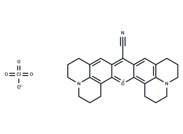
| Formula | C26H26ClN3O5 |
| Cas No. | 137993-41-0 |
| Smiles | [O-][Cl](=O)(=O)=O.N#Cc1c2cc3CCCN4CCCc(c34)c2[o+]c2c3CCCN4CCCc(cc12)c34 |
| Relative Density. | no data available |
| Storage | Keep away from direct sunlight Powder: -20°C for 3 years | In solvent: -80°C for 1 year Shipping with blue ice/Shipping at ambient temperature. | |||||||||||||||||||||||||||||||||||
| Solubility Information | DMSO: 83.33 mg/mL (168.02 mM), Sonication is recommended. | |||||||||||||||||||||||||||||||||||
Solution Preparation Table | ||||||||||||||||||||||||||||||||||||
DMSO
Note : The dilution table applies only to solid products. For liquid products, please calculate the stock solution based on the stated concentration and/or density. | ||||||||||||||||||||||||||||||||||||
 For example, if the intended dosage is 10 mg/kg for animals weighing 20 g , with a dosing volume of 100 μL per animal, and a total of 10 animals are to be administered, using a formulation of
  10% DMSO+ 40% PEG300+ 5% Tween 80+ 45% Saline/PBS/ddH2O , the resulting working solution concentration would be 2 mg/mL.
Dissolve 2 mg of the compound in 100 μL DMSO to obtain a stock solution at a concentration of 20 mg/mL . If the required concentration exceeds the compound's known solubility, please contact us for technical support before proceeding.
1) Add 100 μL of the DMSO stock solution to 400 µL PEG300 and mix thoroughly until the solution becomes clear.
2) Add 50 µL Tween 80 and mix well until fully clarified.
3) Add 450 µL Saline,PBS or ddH2O and mix thoroughly until a homogeneous solution is obtained.
| Size | Quantity | Unit Price | Amount | Operation |
|---|
 Hello! How can I help you today?
 Copyright © 2015-2026 TargetMol Chemicals Inc. All Rights Reserved.
