
Shopping Cart
Remove All Your shopping cart is currently empty


Synonyms: Crystal Violet Stain Solution (0.5%)
| Pack Size | Price | USA Stock | Global Stock | Quantity |
|---|---|---|---|---|
| 100 mL | $41 | In Stock | In Stock |
| Crystal Violet Stain Solution | Features |
|---|---|
| Ingredient | Crystal Violet |
| CAS | 548-62-9 |
| Conc. | 0.5% |
| Solvent | Ethanol |
1.Strong specificity, with high affinity for certain structures (e.g., bacterial peptidoglycan).
2.Broad applicability, suitable for various sample types including bacteria, fungi, plant and animal cells, and tissues.
3.High reagent stability, resistant to degradation, ideal for long-term experimental use.
4.Easy to use.
5.Cost-effective.
1.Gram staining
2.Preliminary histopathological screening
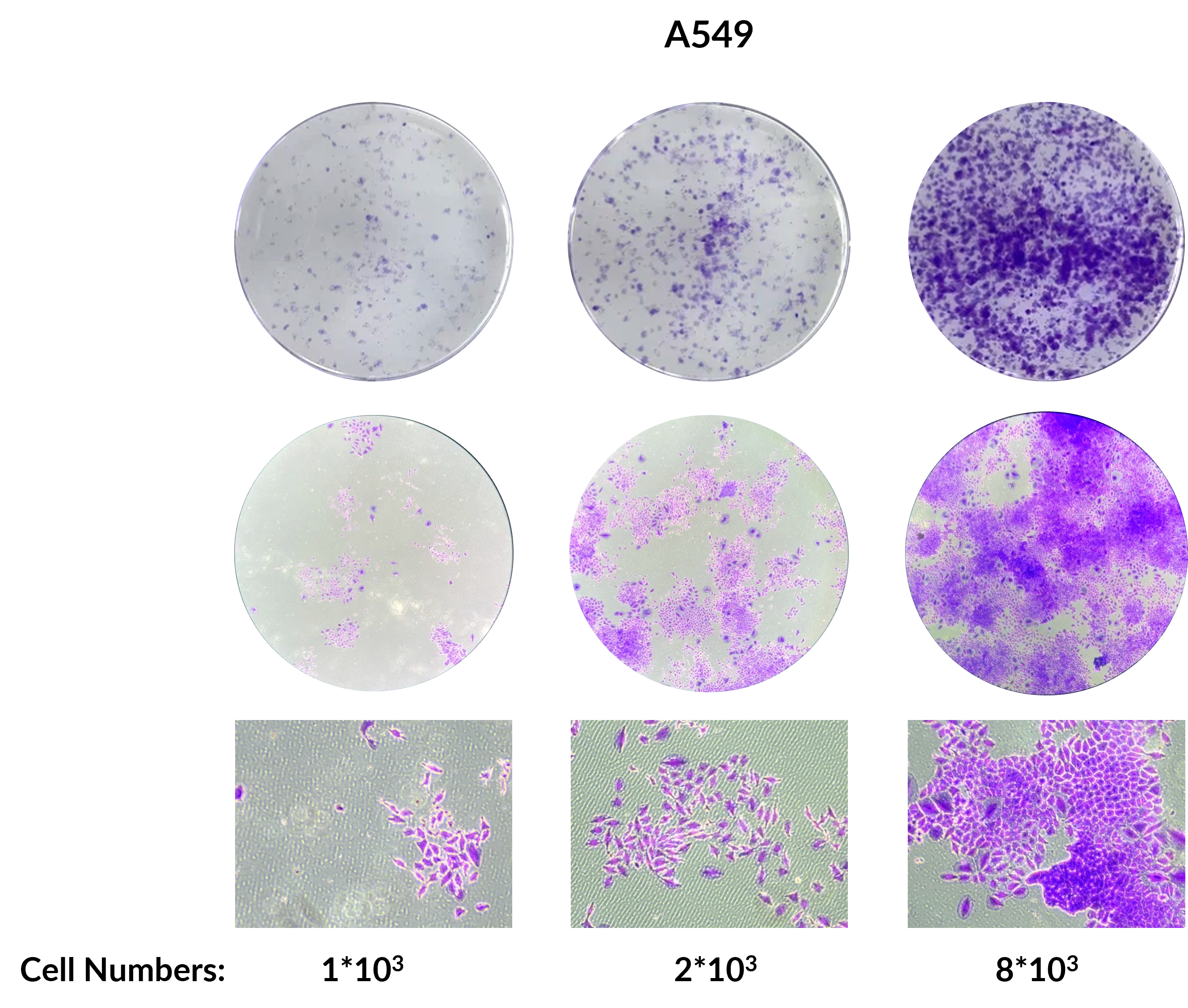
3.Colony formation assay
4.Cell adhesion assay
5.Transwell migration or invasion assay
Dilute the crystal violet stock solution with ddH2O to 0.1% to obtain the staining working solution.
1. Sample Preparation
(1) Paraffin sections: Dewax in xylene for 5-10 min, then replace with fresh xylene and dewax again for 5-10 min. After dewaxing, immerse sequentially in 100% ethanol for 5 min, 90% ethanol for 2 min, and 70% ethanol for 2 min. Finally, wash with ddH₂O for 2 min.
(2) Adherent cells: Fix with 4% paraformaldehyde at 37℃ for 20-30 min, then wash once with PBS buffer.
2. Crystal Violet Staining
(1) Add crystal violet working solution and stain at room temperature for 10-30 min (ensure the staining solution fully covers the sample).
(2) Discard the staining solution and wash with a gentle stream of water until the rinsing water is free of dye (avoid directing the water stream directly onto cells or sections).
(3) Air-dry at room temperature.
3. Observation
Examine and photograph under a microscope.
Store at room temperature, protected from light. Valid for one year.
1.If the experiment involves decolorization (e.g., ethanol decolorization in Gram staining), the decolorization time should be controlled (generally 10-20 seconds) until the effluent is nearly colorless. Over-decolorization may lead to Gram-positive bacteria being misidentified as Gram-negative.
2.It is recommended to perform a preliminary test when using this product for the first time to determine the appropriate staining time.
3.Crystal violet is harmful to humans. Handle with care and take proper protective measures to avoid direct contact with skin or inhalation.
4.The product is for R&D use only, not for diagnostic procedures, food, drug, household or other uses.
5.Please wear a lab coat and disposable gloves.
| Size | Quantity | Unit Price | Amount | Operation |
|---|
 Hello! How can I help you today?
 Copyright © 2015-2026 TargetMol Chemicals Inc. All Rights Reserved.
