
Shopping Cart
Remove All Your shopping cart is currently empty

Synonyms: IR 780 iodide

| Pack Size | Price | USA Stock | Global Stock | Quantity |
|---|---|---|---|---|
| 25 mg | $35 | In Stock | In Stock | |
| 50 mg | $44 | In Stock | In Stock | |
| 100 mg | $59 | In Stock | In Stock | |
| 200 mg | $67 | - | In Stock | |
| 1 mL x 10 mM (in DMSO) | $30 | In Stock | In Stock |
| Description | IR-780 Iodide (IR 780 iodide), a near‐infrared fluorescent dye, is used for the exclusive characterization of human CSCs through the HIF‐1α/glycolysis dependent mitochondrial transporter ABCB10's activity. |
| Synonyms | IR 780 iodide |
| Cell Research | 1. For cancer stem cell (CSC) research 1. Cell staining: Dissolve IR-780 Iodide in an appropriate solvent (such as PBS or DMEM culture medium), usually at a concentration of 10-20 μM. Add the dye solution to the cell culture medium and incubate for 30-60 minutes. 2. Washing: After staining, gently wash the cells with PBS to remove unbound dye. 3. Fluorescence microscopy: Observe using a near-infrared fluorescence microscope (e.g., excitation wavelength 745 nm, emission wavelength 780 nm). IR-780 Iodide can be localized within the cell and its distribution in different subcellular structures, especially mitochondria and cell membrane regions, can be observed. 2. For HIF-1α pathway research 1. Cell treatment: Select a suitable cancer cell line or CSC, add IR-780 Iodide and incubate, and observe the adsorption of the dye and its correlation with mitochondrial transport proteins. 2. Fluorescence detection: Fluorescence imaging technology is used to detect the effect of changes in HIF-1α pathway activity on the distribution and metabolic pathway of IR-780 dye. III. Used for multidrug resistance research 1. Staining and treatment: Add IR-780 Iodide dye to drug-treated cells to study the effect of drugs on the distribution of intracellular dye. 2. Flow cytometry detection: Use flow cytometry to analyze cells, study the binding effects of different drugs and IR-780, and evaluate the activity of transporters. IV. Cell imaging and positioning 1. Staining steps: Add IR-780 Iodide solution to cell culture medium and incubate for a certain period of time (usually 30 minutes to 1 hour). 2. Microscopic imaging: Use a high-resolution near-infrared fluorescence microscope to observe cell staining and fluorescence distribution. The above information is based on published literature. Experimental procedures should be appropriately modified to meet specific research demands. |
| Molecular Weight | 667.12 |
| Formula | C36H44ClIN2 |
| Cas No. | 207399-07-3 |
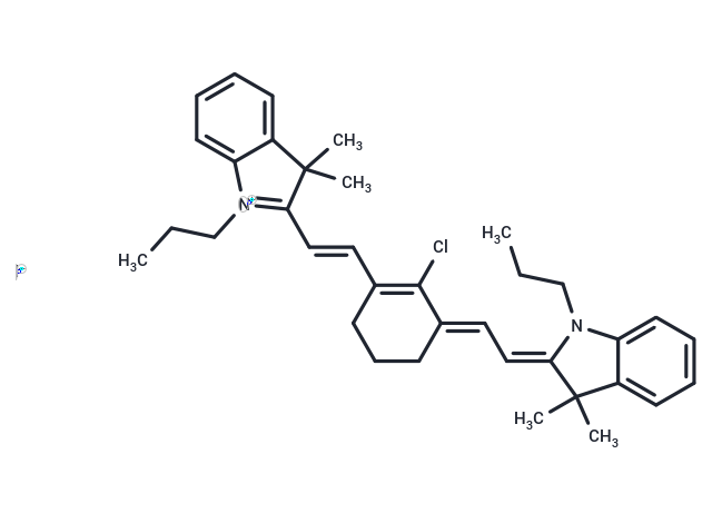
| Smiles | [I-].CCCN1\C(=C/C=C2\CCCC(\C=C\C3=[N+](CCC)c4ccccc4C3(C)C)=C2Cl)C(C)(C)c2ccccc12 |
| Relative Density. | no data available |
| Storage | Keep away from direct sunlight,Store under nitrogen Powder: -20°C for 3 years | In solvent: -80°C for 1 year Shipping with blue ice/Shipping at ambient temperature. | |||||||||||||||||||||||||
| Solubility Information | DMSO: 6.88 mg/mL (10.31 mM), Sonication is recommended. Methanol: 6.25 mg/mL (9.37 mM), Sonication is recommended. | |||||||||||||||||||||||||
Solution Preparation Table | ||||||||||||||||||||||||||
Methanol/DMSO
DMSO
Note : The dilution table applies only to solid products. For liquid products, please calculate the stock solution based on the stated concentration and/or density. | ||||||||||||||||||||||||||
 For example, if the intended dosage is 10 mg/kg for animals weighing 20 g , with a dosing volume of 100 μL per animal, and a total of 10 animals are to be administered, using a formulation of
  10% DMSO+ 40% PEG300+ 5% Tween 80+ 45% Saline/PBS/ddH2O , the resulting working solution concentration would be 2 mg/mL.
Dissolve 2 mg of the compound in 100 μL DMSO to obtain a stock solution at a concentration of 20 mg/mL . If the required concentration exceeds the compound's known solubility, please contact us for technical support before proceeding.
1) Add 100 μL of the DMSO stock solution to 400 µL PEG300 and mix thoroughly until the solution becomes clear.
2) Add 50 µL Tween 80 and mix well until fully clarified.
3) Add 450 µL Saline,PBS or ddH2O and mix thoroughly until a homogeneous solution is obtained.
| Size | Quantity | Unit Price | Amount | Operation |
|---|
 Hello! How can I help you today?
 Copyright © 2015-2026 TargetMol Chemicals Inc. All Rights Reserved.
