
Shopping Cart
Remove All Your shopping cart is currently empty

Synonyms: PTK 2, Protein-tyrosine kinase 2, Protein phosphatase 1 regulatory subunit 71, PPP1R71, pp125FAK, p125FAK, FRNK, Focal adhesion kinase-related nonkinase, Focal adhesion kinase 1, FAK1, FAK, FADK 1, EC 2.7.10.2
Anti-PTK2 Antibody
(4L758)
| Pack Size | Price | USA Stock | Global Stock | Quantity |
|---|---|---|---|---|
| 50 µL | $297 | 7-10 days | 7-10 days | |
| 100 µL | $498 | 7-10 days | 7-10 days |
| Description | Anti-PTK2 Antibody (4L758) is a Rabbit antibody targeting PTK2. Anti-PTK2 Antibody (4L758) can be used in FCM,ICC/IF,IHC,IP,WB. |
| Synonyms | PTK 2, Protein-tyrosine kinase 2, Protein phosphatase 1 regulatory subunit 71, PPP1R71, pp125FAK, p125FAK, FRNK, Focal adhesion kinase-related nonkinase, Focal adhesion kinase 1, FAK1, FAK, FADK 1, EC 2.7.10.2 |
| Ig Type | IgG |
| Clone | 4L758 |
| Reactivity | Human,Mouse,Rat |
| Verified Activity | 1. Western blot analysis of FAK on different lysates using anti-FAK antibody at 1/1,000 dilution. Positive control: Lane 1: Hela, Lane 2: Mouse spleen. 2. Immunohistochemical analysis of paraffin-embedded huamn liver cancer tissue using anti-FAK antibody. Counter stained with hematoxylin. 3. Immunohistochemical analysis of paraffin-embedded human spleen tissue using anti-FAK antibody. Counter stained with hematoxylin. 4. Immunohistochemical analysis of paraffin-embedded mouse brain tissue using anti-FAK antibody. Counter stained with hematoxylin. 5. Immunohistochemical analysis of paraffin-embedded mouse spleen tissue using anti-FAK antibody. Counter stained with hematoxylin. 6. ICC staining FAK in PANC-1 cells (green). The nuclear counter stain is DAPI (blue). Cells were fixed in paraformaldehyde, permeabilised with 0.25% Triton X100/PBS. 7. ICC staining FAK in SH-SY-5Y cells (green). The nuclear counter stain is DAPI (blue). Cells were fixed in paraformaldehyde, permeabilised with 0.25% Triton X100/PBS. 8. Flow cytometric analysis of Hela cells with FAK antibody at 1/50 dilution (blue) compared with an unlabelled control (cells without incubation with primary antibody; red). Alexa Fluor 488-conjugated goat anti rabbit IgG was used as the secondary antibody. 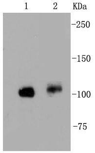 , ,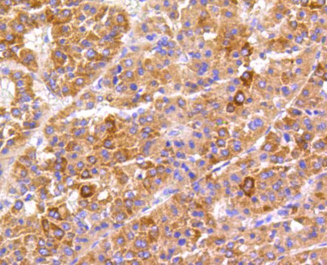 , ,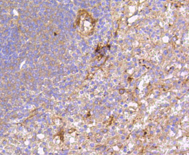 , ,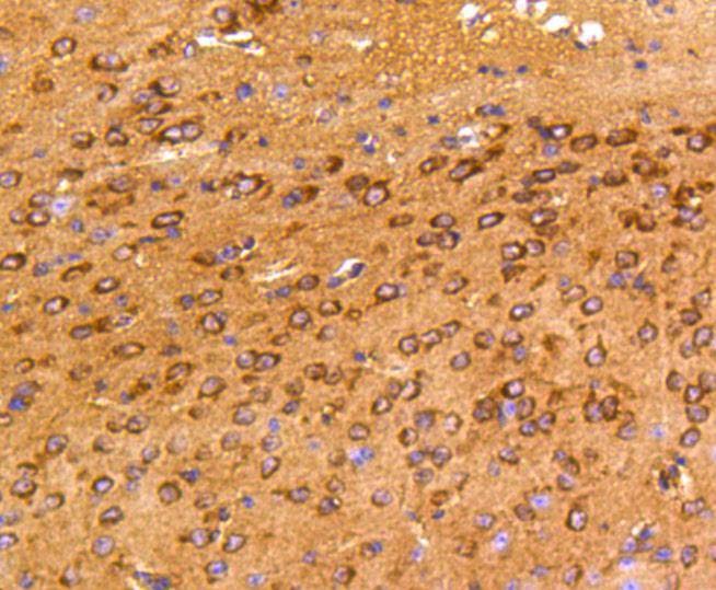 , ,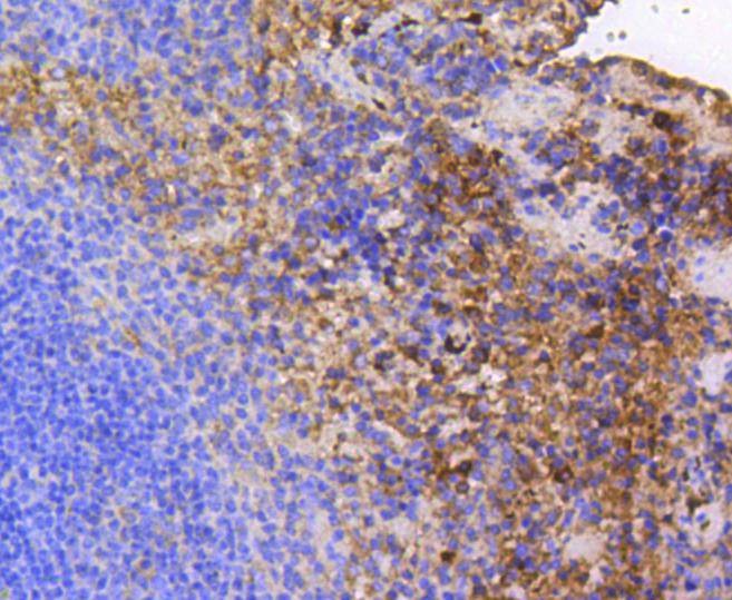 , ,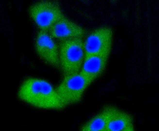 , ,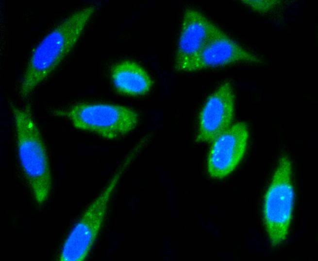 , ,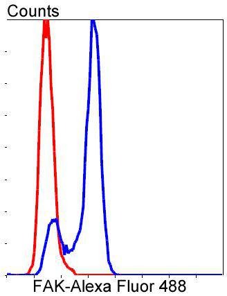 |
| Application | |
| Recommended Dose | WB: 1:1000-2000; IHC: 1:50-200; ICC/IF: 1:50-200; FCM: 1:50-100 |
| Antibody Type | Monoclonal |
| Host Species | Rabbit |
| Construction | Recombinant Antibody |
| Purification | ProA affinity purified |
| Appearance | Liquid |
| Formulation | 1*TBS (pH7.4), 1%BSA, 40%Glycerol. Preservative: 0.05% Sodium Azide. |
| Research Background | Focal adhesion kinase was initially identified as a major substrate for the intrinsic protein tyrosine kinase activity of Src encoded pp60. The deduced amino acid sequence of FAK p125 has shown it to be a cytoplasmic protein tyrosine kinase whose sequence and structural organization are unique as compared to other proteins described to date. Localization of p125 by immunofluorescence suggests that it is primarily found in cellular focal adhesions leading to its designation as focal adhesion kinase (FAK). FAK is concentrated at the basal edge of only those basal keratinocytes that are actively migrating and rapidly proliferating in repairing burn wounds and is activated and localized to the focal adhesions of spreading keratinocytes in culture. Thus, it has been postulated that FAK may have an important in vivo role in the reepithelialization of human wounds. FAK protein tyrosine kinase activity has also been shown to increase in cells stimulated to grow by use of mitogenic neuropeptides or neurotransmitters acting through G protein coupled receptors. |
| Conjucates | Unconjugated |
| Immunogen | Recombinant Protein |
| Uniprot ID |
| Molecular Weight | Theoretical: 119 kDa. |
| Stability & Storage | Store at -20°C or -80°C for 12 months. Avoid repeated freeze-thaw cycles. |
| Transport | Shipping with blue ice. |
| Size | Quantity | Unit Price | Amount | Operation |
|---|
 Hello! How can I help you today?
 Copyright © 2015-2026 TargetMol Chemicals Inc. All Rights Reserved.
