
Shopping Cart
Remove All Your shopping cart is currently empty

Synonyms: T-cell transcription factor NFAT1, T cell transcription factor NFAT 1, Preexisting nuclear factor of activated T cells 2, preexisting component, Nuclear factor of activated T-cells, Nuclear factor of activated T cells pre-existing component, Nuclear factor of activated T cells cytoplasmic calcineurin dependent 2, Nuclear factor of activated T cells cytoplasmic 2, Nuclear factor of activated T cells, NF-ATp, NFATp, NF-ATc2, NFATc2, NFAT1-D, NFAT1, NFAT transcription complex, NFAT pre-existing subunit, NFAT pre existing subunit, NFAT 1, NFAC2_HUMAN, NF ATp, NF ATc2, KIAA0611, cytoplasmic 2, AI607462
Anti-NFAT1 Antibody
(4I426)
| Pack Size | Price | USA Stock | Global Stock | Quantity |
|---|---|---|---|---|
| 50 µL | $296 | 7-10 days | 7-10 days | |
| 100 µL | $497 | 7-10 days | 7-10 days |
| Description | Anti-NFAT1 Antibody (4I426) is a Rabbit antibody targeting NFAT1. Anti-NFAT1 Antibody (4I426) can be used in FCM,ICC/IF,IHC,WB. |
| Synonyms | T-cell transcription factor NFAT1, T cell transcription factor NFAT 1, Preexisting nuclear factor of activated T cells 2, preexisting component, Nuclear factor of activated T-cells, Nuclear factor of activated T cells pre-existing component, Nuclear factor of activated T cells cytoplasmic calcineurin dependent 2, Nuclear factor of activated T cells cytoplasmic 2, Nuclear factor of activated T cells, NF-ATp, NFATp, NF-ATc2, NFATc2, NFAT1-D, NFAT1, NFAT transcription complex, NFAT pre-existing subunit, NFAT pre existing subunit, NFAT 1, NFAC2_HUMAN, NF ATp, NF ATc2, KIAA0611, cytoplasmic 2, AI607462 |
| Ig Type | IgG |
| Clone | 4I426 |
| Reactivity | Human |
| Verified Activity | 1. Immunohistochemical analysis of paraffin-embedded human spleen tissue using anti-NFAT1 antibody. Counter stained with hematoxylin. 2. Immunohistochemical analysis of paraffin-embedded human tonsil tissue using anti-NFAT1 antibody. Counter stained with hematoxylin. 3. Immunohistochemical analysis of paraffin-embedded human colon cancer tissue using anti-NFAT1 antibody. Counter stained with hematoxylin. 4. ICC staining NFAT1 in MCF-7 cells (red). The nuclear counter stain is DAPI (blue). Cells were fixed in paraformaldehyde, permeabilised with 0.25% Triton X100/PBS. 5. ICC staining NFAT1 in Hela cells (red). The nuclear counter stain is DAPI (blue). Cells were fixed in paraformaldehyde, permeabilised with 0.25% Triton X100/PBS. 6. ICC staining NFAT1 in SHG-44 cells (red). The nuclear counter stain is DAPI (blue). Cells were fixed in paraformaldehyde, permeabilised with 0.25% Triton X100/PBS. 7. Flow cytometric analysis of Jurkat cells with NFAT1 antibody at 1/50 dilution (red) compared with an unlabelled control (cells without incubation with primary antibody; black). Alexa Fluor 488-conjugated goat anti rabbit IgG was used as the secondary antibody. 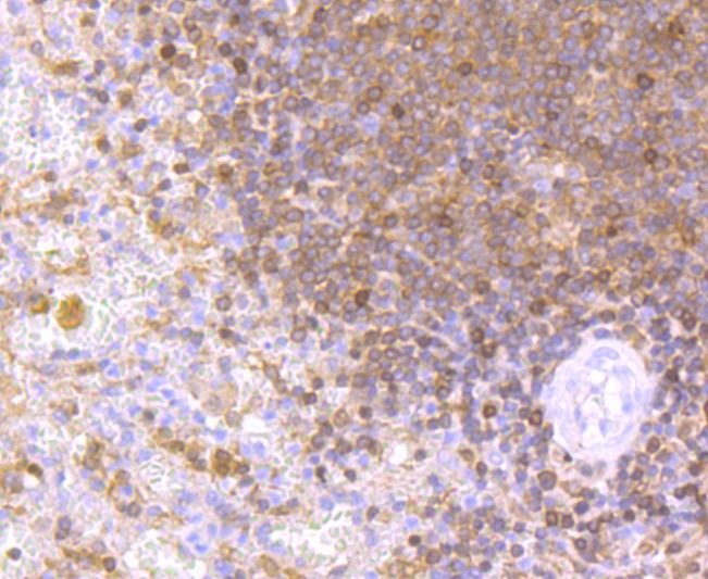 , ,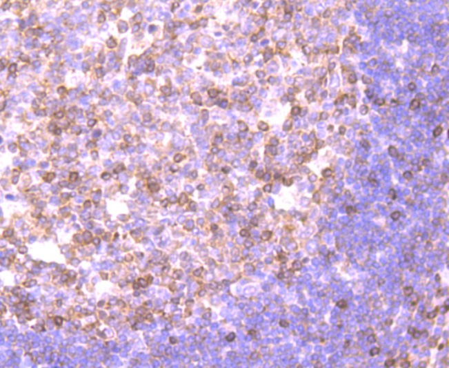 , ,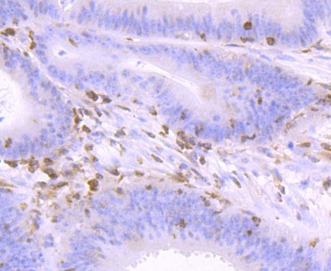 , ,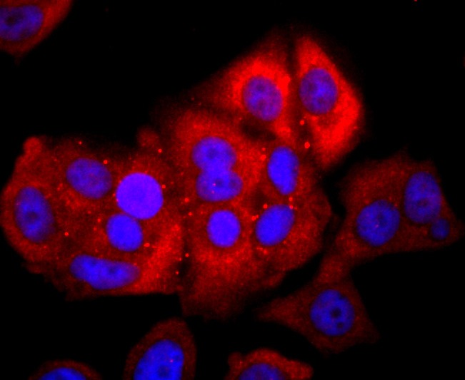 , ,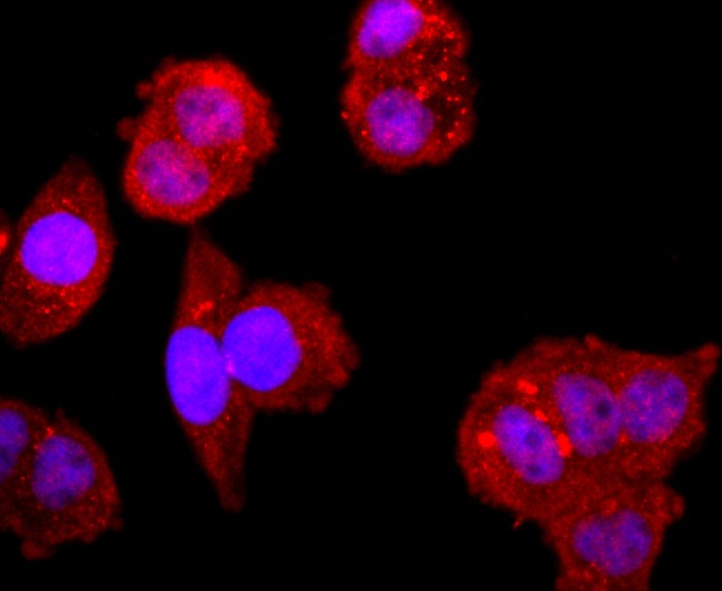 , ,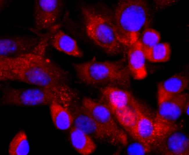 , ,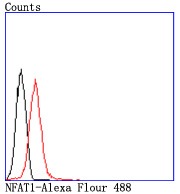 |
| Application | |
| Recommended Dose | WB: 1:500; IHC: 1:50-200; ICC/IF: 1:50-200; FCM: 1:50-100 |
| Antibody Type | Monoclonal |
| Host Species | Rabbit |
| Construction | Recombinant Antibody |
| Purification | ProA affinity purified |
| Appearance | Liquid |
| Formulation | 1*TBS (pH7.4), 1%BSA, 40%Glycerol. Preservative: 0.05% Sodium Azide. |
| Research Background | The NFAT (nuclear factor of activated T cells) family of transcription factors regulates cytokine expression in T cells. Members of the family include NFATc1 (NFATc), NFATc2 (NFATp), NFATn, NFATc3 (NFAT4, NFATx) and NFATc4 (NFAT3). Recognition of antigen by the T cell receptor (TCR) eventually activates the calcium-dependent protein phosphatase calcineurin. Once activated, calcineurin stimulates the translocation of NFATc1 (cytoplasmic) from the cytoplasm to the nucleus where it associates with NFATn (nuclear). Like NFATc1, NFATc2 resides in the cytoplasm and translocates to the nucleus subsequent to activation of calcineurin. Once in the nucleus, NFATc2 synergizes with AP-1 transcription factors to initiate transcription of cytokine genes. NFATc3 and NFATc4 share 65% sequence identity with other members of the NFAT family. They are similar to NFATc2 in that they also synergize with the AP-1 family of proteins. |
| Conjucates | Unconjugated |
| Immunogen | Recombinant Protein |
| Uniprot ID |
| Molecular Weight | Theoretical: 135 kDa. |
| Stability & Storage | Store at -20°C or -80°C for 12 months. Avoid repeated freeze-thaw cycles. |
| Transport | Shipping with blue ice. |
| Size | Quantity | Unit Price | Amount | Operation |
|---|
 Hello! How can I help you today?
 Copyright © 2015-2026 TargetMol Chemicals Inc. All Rights Reserved.
