
Shopping Cart
Remove All Your shopping cart is currently empty

Synonyms: LAB7, CD80 molecule, CD80, CD28LG1, BB1, B7-1, B71, B7.1, B7
Anti-CD80 Polyclonal Antibody 2
| Pack Size | Price | USA Stock | Global Stock | Quantity |
|---|---|---|---|---|
| 50 µL | $220 | 7-10 days | 7-10 days | |
| 100 µL | $373 | 7-10 days | 7-10 days | |
| 200 µL | $528 | 7-10 days | 7-10 days |
| Description | Anti-CD80 Polyclonal Antibody 2 is a Rabbit antibody targeting CD80. Anti-CD80 Polyclonal Antibody 2 can be used in FCM,WB. |
| Synonyms | LAB7, CD80 molecule, CD80, CD28LG1, BB1, B7-1, B71, B7.1, B7 |
| Ig Type | IgG |
| Reactivity | Human,Mouse,Rat |
| Verified Activity | 1. Sample: Lane 1: Raw264.7 (Mouse) Cell Lysate at 30 μg Lane 2: SP2/0 (Mouse) Cell Lysate at 30 μg Lane 3: Thymus (Rat) Lysate at 40 μg Lane 4: Raji (Human) Cell Lysate at 30 μg Lane 5: K562 (Human) Cell Lysate at 30 μg Primary: Anti-CD80 (TMAB-00393) at 1/1000 dilution Secondary: IRDye800CW Goat Anti-Rabbit IgG at 1/20000 dilution Predicted band size: 30 kDa Observed band size: 60 kDa 2. Blank control: Raji. Primary Antibody (green line): Rabbit Anti-CD80 antibody (TMAB-00393) Dilution: 2 μg/Test; Secondary Antibody: Goat anti-rabbit IgG-AF488 Dilution: 0.5 μg/Test. Protocol The cells were incubated in 5% BSA to block non-specific protein-protein interactions for 30 min at room temperature. Cells stained with Primary Antibody for 30 min at room temperature. The secondary antibody used for 40 min at room temperature. 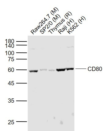 , ,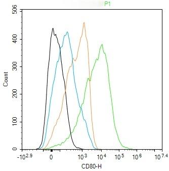 |
| Application | |
| Recommended Dose | WB: 1:500-2000; FCM: 2µg/Test |
| Antibody Type | Polyclonal |
| Host Species | Rabbit |
| Subcellular Localization | Membrane; Single-pass type I membrane protein. |
| Tissue Specificity | Expressed on activated B-cells, macrophages and dendritic cells. |
| Construction | Polyclonal Antibody |
| Purification | Protein A purified |
| Appearance | Liquid |
| Formulation | 0.01M TBS (pH7.4) with 1% BSA, 0.02% Proclin300 and 50% Glycerol. |
| Concentration | 1 mg/mL |
| Research Background | CD80 is a member of the Ig superfamily and serves as the ligand for two T cell molecules, CD28 and CTLA4. Interactions between CD28 and CD80 on activated B cells result in enhanced T cell activation. CD80 is rapidly induced on the surface of in vitro activated B cells, Epstein Barr Virus (EBV) transformed B cell lines, Burkitts lymphoma cell lines, freshly isolated follicular B lymphoma cells, T cells, and monocytes. It is also expressed at high levels in dendritic cells. It reacts weakly with a small proportion of non activated normal B cells and with HTLV1 infected T cells. CD80 does not react with peripheral monocytes, resting and activated normal T cells, T cell lines and T cell clones, nor with myelomonocytic cell lines. |
| Immunogen | KLH conjugated synthetic peptide: rat CD80 |
| Antigen Species | Rat |
| Gene Name | CD80 |
| Protein Name | T-lymphocyte activation antigen CD80 |
| Biology Area | CD,B Lymphocytic Lineage,Dendritic Cell Lineage,Monocytic Lineage |
| Function | Involved in the costimulatory signal essential for T lymphocytes activation. T-cell proliferation and cytokine production is induced by the binding of CD28 or CTLA-4 to this receptor. |
| Molecular Weight | Theoretical: 30 kDa. Actual: 60 kDa. |
| Stability & Storage | Store at -20°C or -80°C for 12 months. Avoid repeated freeze-thaw cycles. |
| Transport | Shipping with blue ice. |
| Size | Quantity | Unit Price | Amount | Operation |
|---|
 Hello! How can I help you today?
 Copyright © 2015-2026 TargetMol Chemicals Inc. All Rights Reserved.
