
Shopping Cart
Remove All Your shopping cart is currently empty

Synonyms: RNCASP9, OTTHUMP00000044594, MCH6, MCH 6, ICE like apoptotic protease 6, ICE LAP6, Caspase-9 subunit p35, Caspase9, Caspase 9c, Caspase 9 precursor, Caspase 9 apoptosis related cysteine protease, Caspase 9, CASP9, CASP 9, Apoptotic protease MCH6, Apoptotic protease MCH 6, Apoptotic protease activating factor 3, Apoptosis related cysteine peptidase, APAF3, APAF 3
Anti-Caspase-9 Polyclonal Antibody
| Pack Size | Price | USA Stock | Global Stock | Quantity |
|---|---|---|---|---|
| 50 µL | $220 | 7-10 days | 7-10 days | |
| 100 µL | $374 | 7-10 days | 7-10 days | |
| 200 µL | $528 | 7-10 days | 7-10 days |
| Description | Anti-Caspase-9 Polyclonal Antibody is a Rabbit antibody targeting Caspase-9. Anti-Caspase-9 Polyclonal Antibody can be used in FCM, ICC/IF, IF, IHC-Fr, IHC-P, WB. |
| Synonyms | RNCASP9, OTTHUMP00000044594, MCH6, MCH 6, ICE like apoptotic protease 6, ICE LAP6, Caspase-9 subunit p35, Caspase9, Caspase 9c, Caspase 9 precursor, Caspase 9 apoptosis related cysteine protease, Caspase 9, CASP9, CASP 9, Apoptotic protease MCH6, Apoptotic protease MCH 6, Apoptotic protease activating factor 3, Apoptosis related cysteine peptidase, APAF3, APAF 3 |
| Ig Type | IgG |
| Reactivity | Human,Mouse,Rat (predicted:Dog,Cow) |
| Verified Activity | 1. Blank control: RSC96 (blue), the cells were fixed with 2% paraformaldehyde (10 min) and then permeabilized with ice-cold 90% methanol for 30 min on ice. Isotype Control Antibody: Rabbit Igg (orange); Secondary Antibody: Goat anti-rabbit IgG-Pe (white blue), Dilution: 1:200 in 1 X PBS containing 0.5% BSA; Primary Antibody Dilution: 1 μg in 100 μL 1X PBS containing 0.5% BSA (green). 2. Blank control: K562 (blue). Primary Antibody: Rabbit Anti-caspase-9 antibody (TMAB-00292,Green); Dilution: 1 μg in 100 μL 1X PBS containing 0.5% BSA; Isotype Control Antibody: Rabbit Igg (orange),used under the same conditions; Secondary Antibody: Goat anti-rabbit IgG-FITC (white blue), Dilution: 1:200 in 1 X PBS containing 0.5% BSA. Protocol The cells were fixed with 80% methanol (5 min) and and then permeabilized with 0.01M PBS-Tween for 20 min. Primary antibody (TMAB-00292, 1 μg/1x10^6 cells) were incubated for 30 min at room temperature, followed by 1 X PBS containing 0.5% BSA + 10% goat serum (30 min) to block non-specific protein-protein interactions. Then the Goat Anti-rabbit IgG/FITC antibody was added into the blocking buffer mentioned above to react with the primary antibody at 1/200 dilution for 30 min at room temperature. 3. Paraformaldehyde-fixed, paraffin embedded (rat lung); Antigen retrieval by boiling in sodium citrate buffer (pH6.0) for 15 min; Block endogenous peroxidase by 3% hydrogen peroxide for 20 min; Blocking buffer (normal goat serum) at 37°C for 30 min; Antibody incubation with (Insulin like growth factor 1) Polyclonal Antibody, Unconjugated at 1:400 overnight at 4°C, followed by a conjugated secondary antibody for 20 min and DAB staining. 4. Tissue/cell: Hela cell; 4% Paraformaldehyde-fixed; Triton X-100 at room temperature for 20 min; Blocking buffer (normal goat serum) at 37°C for 20 min; Antibody incubation with (Caspase-9) polyclonal Antibody, Unconjugated (TMAB-00292) 1:100, 90 minutes at 37°C; followed by a FITC conjugated Goat Anti-Rabbit IgG antibody at 37°C for 90 minutes, DAPI (blue) was used to stain the cell nucleus. 5. Paraformaldehyde-fixed, paraffin embedded (rat pancreas); Antigen retrieval by boiling in sodium citrate buffer (pH6.0) for 15 min; Block endogenous peroxidase by 3% hydrogen peroxide for 20 min; Blocking buffer (normal goat serum) at 37°C for 30 min; Antibody incubation with (Caspase-9) Polyclonal Antibody, Unconjugated (TMAB-00292) at 1:200 overnight at 4°C, followed by operating according to SP Kit (Rabbit) instructionsand DAB staining. 6. Tissue/cell: HepG2 cell; 4% Paraformaldehyde-fixed; Triton X-100 at room temperature for 20 min; Blocking buffer (normal goat serum) at 37°C for 20 min; Antibody incubation with (Caspase-9) polyclonal Antibody, Unconjugated (TMAB-00292) 1:100, 90 minutes at 37°C; followed by a FITC conjugated Goat Anti-Rabbit IgG antibody at 37°C for 90 minutes, DAPI (blue) was used to stain the cell nucleus. 7. Tissue/cell: MCF-7 cell; 4% Paraformaldehyde-fixed; Triton X-100 at room temperature for 20 min; Blocking buffer (normal goat serum) at 37°C for 20 min; Antibody incubation with (Caspase-9) Polyclonal Antibody, Unconjugated (TMAB-00292) 1:50, 90 minutes at 37°C; followed by a conjugated Goat Anti-Rabbit IgG antibody at 37°C for 90 minutes, DAPI (blue) was used to stain the cell nucleus. 8. HepG2 cell; 4% Paraformaldehyde-fixed; Triton X-100 at room temperature for 20 min; Blocking buffer (normal goat serum) at 37°C for 20 min; Antibody incubation with (Caspase-9) polyclonal Antibody, Unconjugated (TMAB-00292) 1:100, 90 minutes at 37°C; followed by a conjugated Goat Anti-Rabbit IgG antibody at 37°C for 90 minutes, DAPI (blue) was used to stain the cell nucleus. 9. Sample: 293T-UV Cell (Human) Lysate at 30 μg Primary: Anti-Caspase-9 at 1/300 dilution Secondary: IRDye800CW Goat Anti-Rabbit IgG at 1/20000 dilution Predicted band size: 35/50 kDa Observed band size: 35/50 kDa 10. Sample: Lane 1: Mouse Stomach tissue lysates Lane 2: Mouse Spleen tissue lysates Primary: Anti-Caspase-9 (TMAB-00292) at 1/1000 dilution Secondary: IRDye800CW Goat Anti-Rabbit IgG at 1/20000 dilution Predicted band size: 35/50 kDa Observed band size: 52 kDa 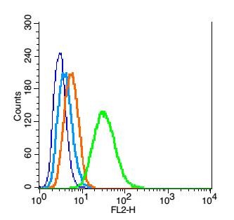 , ,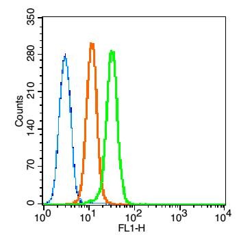 , ,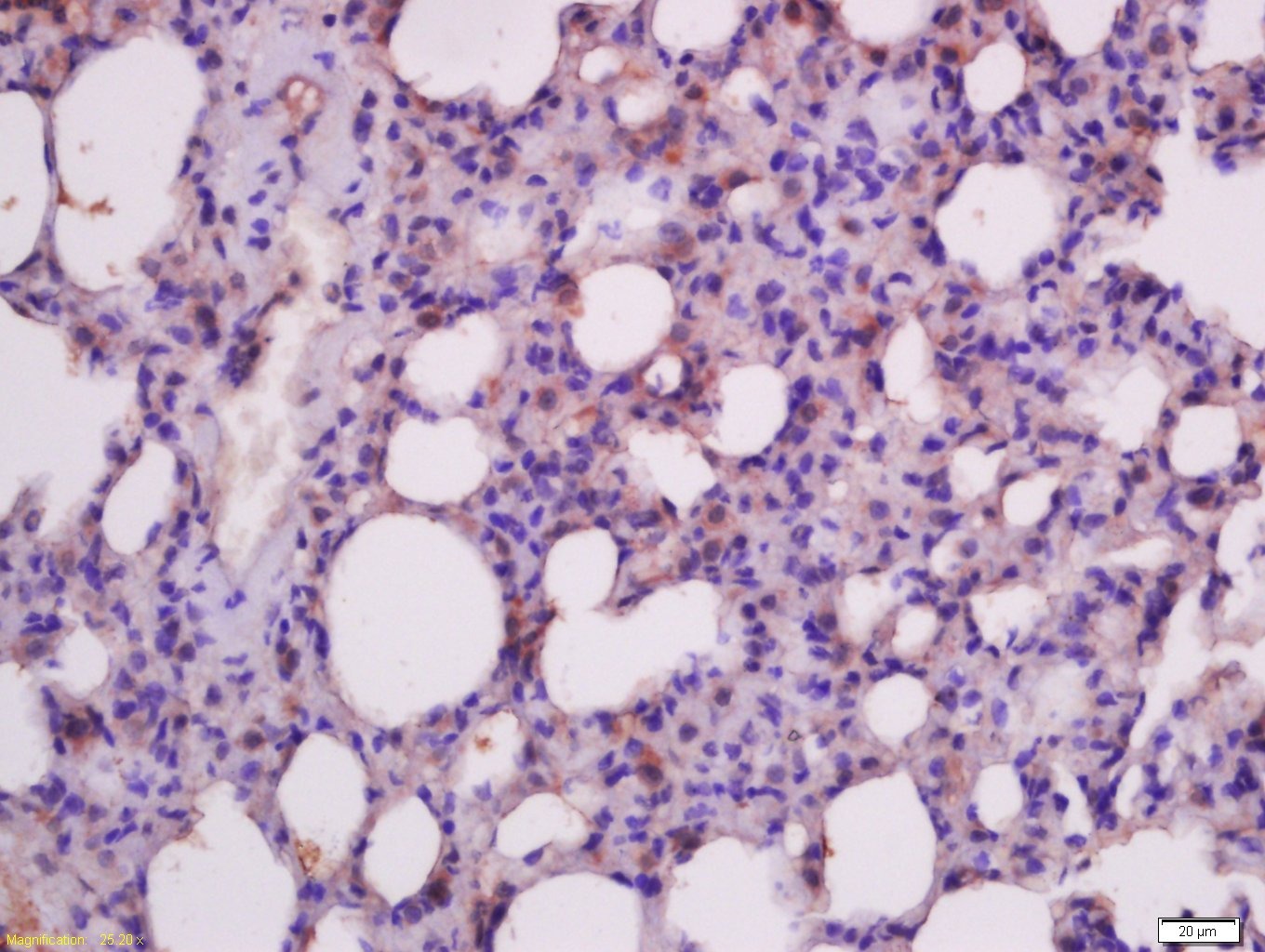 , ,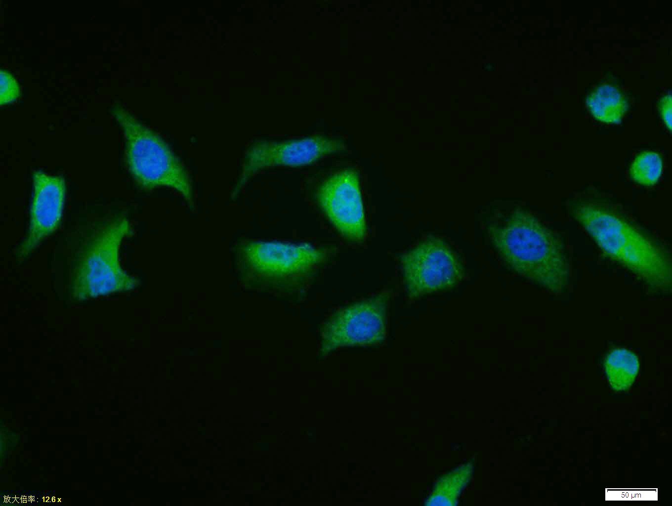 , ,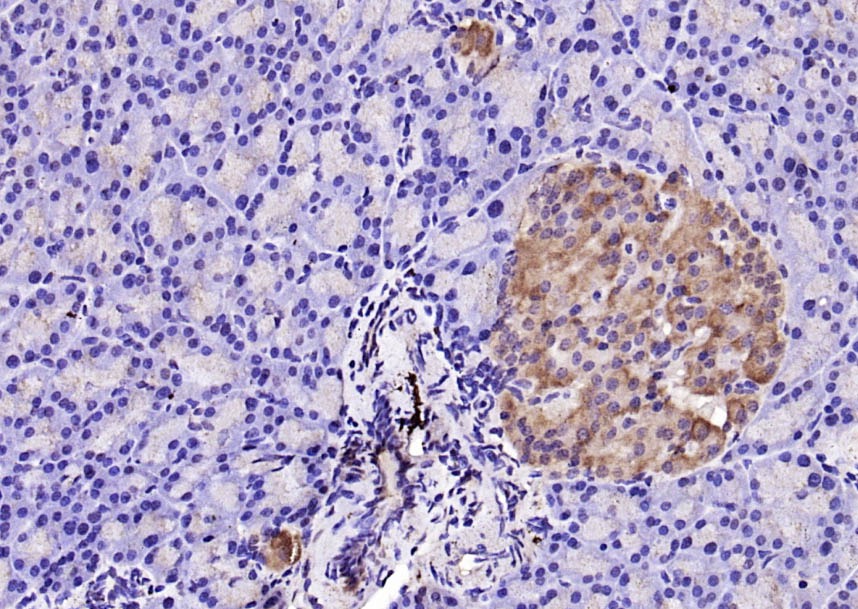 , ,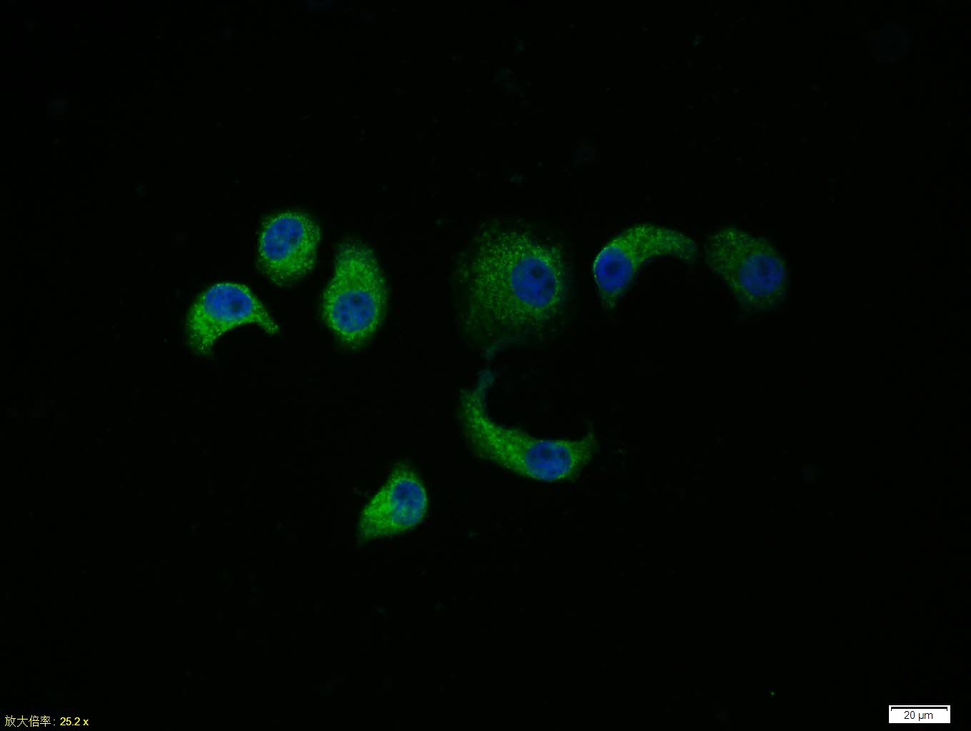 , ,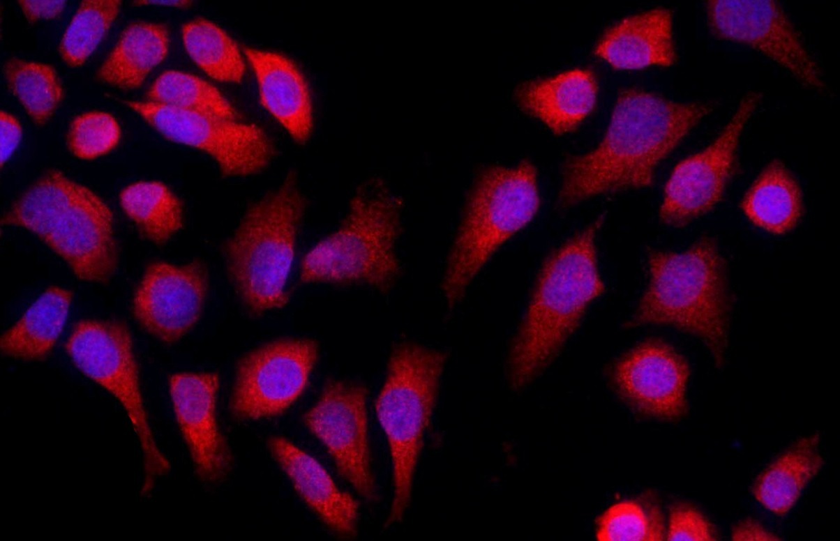 , ,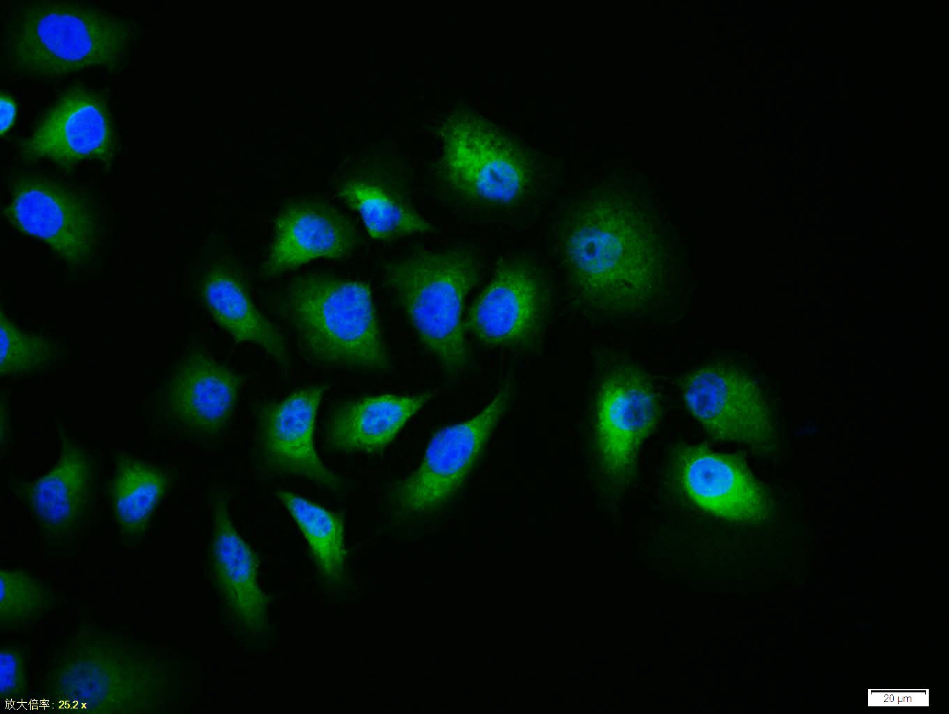 , ,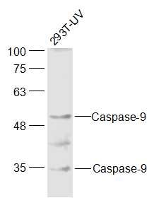 , ,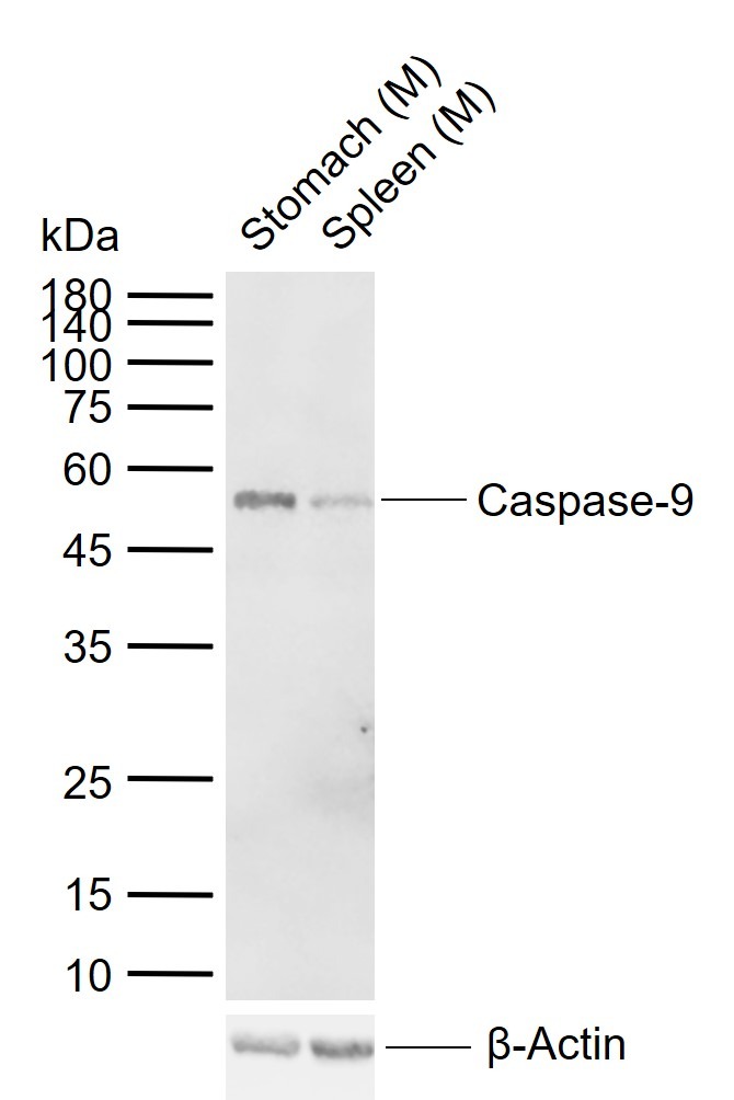 |
| Application | |
| Recommended Dose | FCM=1 μg/Test; ICC/IF=1:100-500; IF=1:100-500; IHC-Fr=1:100-500; IHC-P=1:100-500; WB=1:500-2000 |
| Antibody Type | Polyclonal |
| Host Species | Rabbit |
| Tissue Specificity | Ubiquitous, with highest expression in the heart, moderate expression in liver, skeletal muscle, and pancreas. Low levels in all other tissues. Within the heart, specifically expressed in myocytes. |
| Construction | Polyclonal Antibody |
| Purification | Protein A purified |
| Appearance | Liquid |
| Formulation | 0.01M TBS (pH7.4) with 1% BSA, 0.02% Proclin300 and 50% Glycerol. |
| Concentration | 1 mg/mL |
| Research Background | Caspase 9 (also known as ICE like apoptotic protease 6 (ICE LAP6), apoptotic protease Mch6, and apoptotic protease activating factor 3 (Apaf3)) is a member of the peptidase family C14 that contains a CARD domain. This caspase is active as a heterotetramer and has been reported to have two isoforms. ProCaspase 9 has been reported to be approximately 47 kD. This caspase is present in the cytosol and, upon activation, translocates to the mitochondria. Caspase 9 is involved in the caspase activation cascade responsible for apoptosis execution and cleaves/activates Caspase 3 and Caspase 6. Caspase 9 is inhibited by the dominant negative isoform, BclXL, cIAP1, cIAP2, XIAP, and Livin. This caspase becomes activated when recruited to Apaf1/cytochrome c complex, and following cleavage by Apaf1, granzyme B, Caspase 3, possibly Caspase 8 and Caspase 10 into large p37 and small p10 subunits. Caspase 9 intereacts with BIRC7 and has been shown to cleave PARP and vimentin. |
| Immunogen | KLH conjugated synthetic peptide: human Caspase-9 subunit p35 |
| Antigen Species | Human |
| Gene Name | CASP9 |
| Gene ID | |
| Protein Name | Caspase-9 |
| Uniprot ID | |
| Biology Area | Caspases,Cytochrome C,Metabolism,Caspases,Cytochrome C,Caspases,Apoptosis |
| Function | Involved in the activation cascade of caspases responsible for apoptosis execution. Binding of caspase-9 to Apaf-1 leads to activation of the protease which then cleaves and activates caspase-3. Proteolytically cleaves poly(ADP-ribose) polymerase (PARP). Isoform 2 lacks activity is an dominant-negative inhibitor of caspase-9. |
| Molecular Weight | Theoretical: 35/50 kDa. Actual: 52 kDa. |
| Stability & Storage | Store at -20°C or -80°C for 12 months. Avoid repeated freeze-thaw cycles. |
| Transport | Shipping with blue ice. |
| Size | Quantity | Unit Price | Amount | Operation |
|---|
 Hello! How can I help you today?
 Copyright © 2015-2026 TargetMol Chemicals Inc. All Rights Reserved.
