
Shopping Cart
Remove All Your shopping cart is currently empty

Synonyms: p45, Interleukin-1 beta-converting enzyme (ICE;IL-1 beta-converting enzyme), Interleukin-1 beta convertase (IL-1BC), IL1BCE, IL1BC, Caspase-1, CASP-1, CASP1
Anti-Caspase-1 p10 Polyclonal Antibody
| Pack Size | Price | USA Stock | Global Stock | Quantity |
|---|---|---|---|---|
| 50 µL | $222 | 7-10 days | 7-10 days | |
| 100 µL | $373 | 7-10 days | 7-10 days | |
| 200 µL | $529 | 7-10 days | 7-10 days |
| Description | Anti-Caspase-1 p10 Polyclonal Antibody is a Rabbit antibody targeting Caspase-1 p10. Anti-Caspase-1 p10 Polyclonal Antibody can be used in FCM,IF,IHC-Fr,IHC-P. |
| Synonyms | p45, Interleukin-1 beta-converting enzyme (ICE;IL-1 beta-converting enzyme), Interleukin-1 beta convertase (IL-1BC), IL1BCE, IL1BC, Caspase-1, CASP-1, CASP1 |
| Ig Type | IgG |
| Reactivity | Human,Mouse,Rat |
| Verified Activity | 1. Tissue/cell: rat transplant lymphoma; 4% Paraformaldehyde-fixed and paraffin-embedded; Antigen retrieval: citrate buffer (0.01M, pH6.0), Boiling bathing for 15 min; Block endogenous peroxidase by 3% Hydrogen peroxide for 30 min; Blocking buffer (normal goat serum) at 37°C for 20 min; Incubation: Anti-Caspase-1 Polyclonal Antibody, Unconjugated (TMAB-00280) 1:200, overnight at 4°C, followed by conjugation to the secondary antibody and DAb staining. 2. Tissue/cell: human lung carcinoma; 4% Paraformaldehyde-fixed and paraffin-embedded; Antigen retrieval: citrate buffer (0.01M, pH6.0), Boiling bathing for 15 min; Block endogenous peroxidase by 3% Hydrogen peroxide for 30 min; Blocking buffer (normal goat serum) at 37°C for 20 min; Incubation: Anti-Caspase-1 Polyclonal Antibody, Unconjugated (TMAB-00280) 1:200, overnight at 4°C, followed by conjugation to the secondary antibody and DAb staining. 3. Paraformaldehyde-fixed, paraffin embedded (Rat brain); Antigen retrieval by boiling in sodium citrate buffer (pH6.0) for 15 min; Block endogenous peroxidase by 3% hydrogen peroxide for 20 min; Blocking buffer (normal goat serum) at 37°C for 30 min; Antibody incubation with (Caspase-1 P10) Polyclonal Antibody, Unconjugated (TMAB-00280) at 1:500 overnight at 4°C, followed by a conjugated secondary for 20 min and DAB staining. 4. Paraformaldehyde-fixed, paraffin embedded (Mouse brain); Antigen retrieval by boiling in sodium citrate buffer (pH6.0) for 15 min; Block endogenous peroxidase by 3% hydrogen peroxide for 20 min; Blocking buffer (normal goat serum) at 37°C for 30 min; Antibody incubation with (Caspase-1 P10) Polyclonal Antibody, Unconjugated (TMAB-00280) at 1:500 overnight at 4°C, followed by a conjugated secondary for 20 min and DAB staining. 5. Blank control: HL-60. Primary Antibody (green line): Rabbit Anti-Caspase-1 P10 antibody (TMAB-00280) Dilution: 1 μg/10^6 cells; Isotype Control Antibody (orange line): Rabbit IgG. Secondary Antibody: Goat anti-rabbit IgG-AF488 Dilution: 1 μg/test. Protocol The cells were fixed with 4% PFA (10 min at room temperature) and then permeabilized with 0.1% PBST for 20 min at room temperature. The cells were then incubated in 5% BSA to block non-specific protein-protein interactions for 30 min at room temperature. Cells stained with Primary Antibody for 30 min at room temperature. The secondary antibody used for 40 min at room temperature. 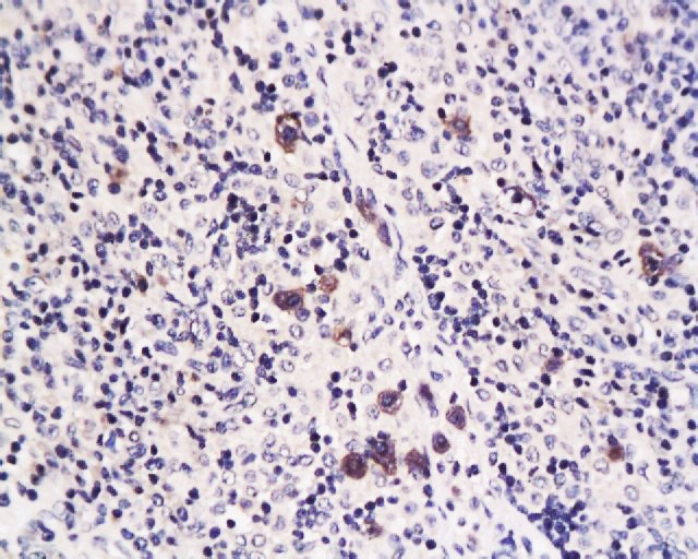 , ,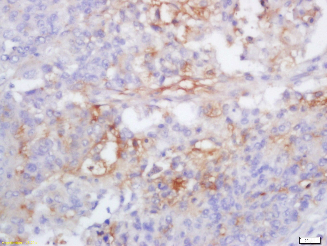 , ,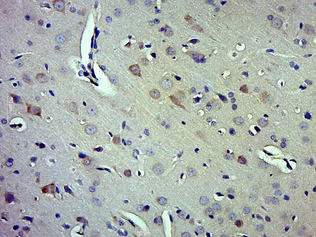 , ,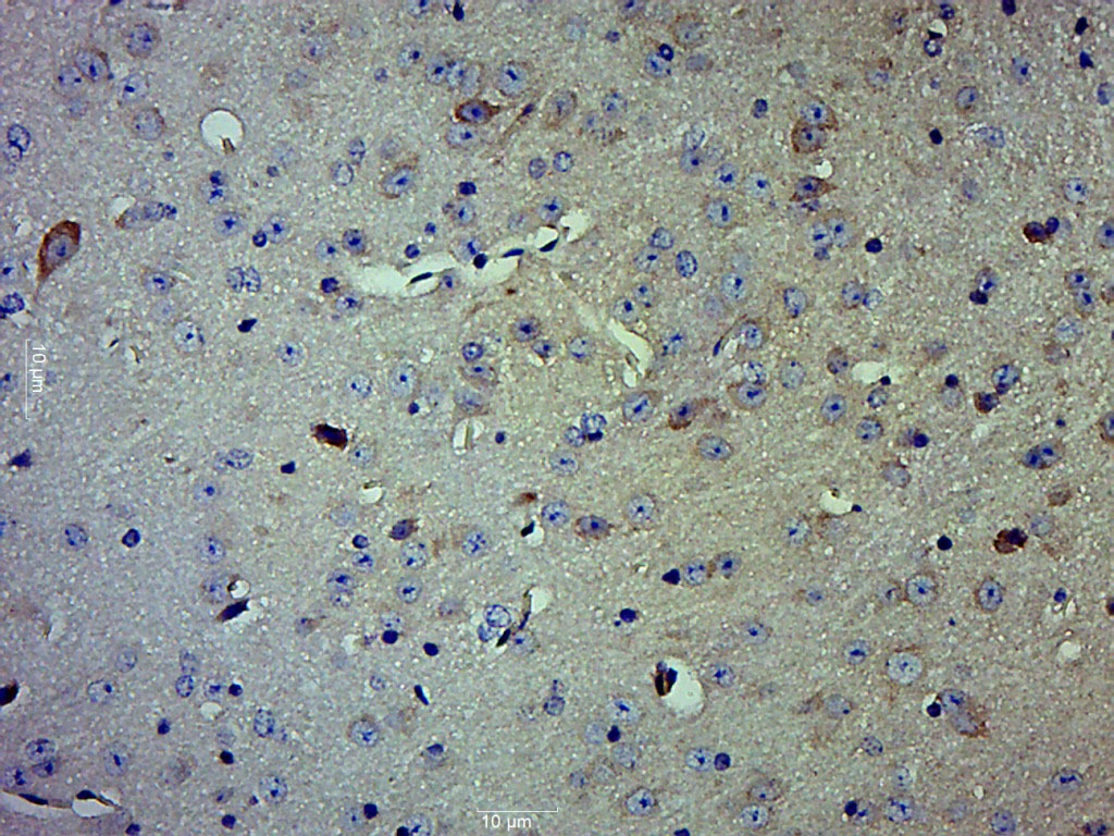 , ,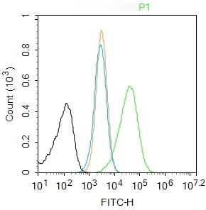 |
| Application | |
| Recommended Dose | IHC-P: 1:100-500; IHC-Fr: 1:100-500; IF: 1:100-500; FCM: 1ug/Test |
| Antibody Type | Polyclonal |
| Host Species | Rabbit |
| Subcellular Localization | Cytoplasm. |
| Tissue Specificity | Expressed in larger amounts in spleen and lung. Detected in liver, heart, small intestine, colon, thymus, prostate, skeletal muscle, peripheral blood leukocytes, kidney and testis. No expression in the brain. |
| Construction | Polyclonal Antibody |
| Purification | Protein A purified |
| Appearance | Liquid |
| Formulation | 0.01M TBS (pH7.4) with 1% BSA, 0.02% Proclin300 and 50% Glycerol. |
| Concentration | 1 mg/mL |
| Research Background | This gene encodes a protein which is a member of the cysteine-aspartic acid protease (caspase) family. Sequential activation of caspases plays a central role in the execution-phase of cell apoptosis. Caspases exist as inactive proenzymes which undergo proteolytic processing at conserved aspartic residues to produce 2 subunits, large and small, that dimerize to form the active enzyme. This gene was identified by its ability to proteolytically cleave and activate the inactive precursor of interleukin-1, a cytokine involved in the processes such as inflammation, septic shock, and wound healing. This gene has been shown to induce cell apoptosis and may function in various developmental stages. Studies of a similar gene in mouse suggest a role in the pathogenesis of Huntington disease. Alternative splicing of this gene results in five transcript variants encoding distinct isoforms. [provided by RefSeq]. |
| Immunogen | KLH conjugated synthetic peptide: human Caspase-1 |
| Antigen Species | Human |
| Gene Name | CASP1 |
| Gene ID | |
| Protein Name | Caspase-1 |
| Uniprot ID | |
| Biology Area | Response to hypoxia,Caspases,Metabolism,Caspases,Caspases,Apoptosis,Hypoxia |
| Function | Thiol protease that cleaves IL-1 beta between an Asp and an Ala, releasing the mature cytokine which is involved in a variety of inflammatory processes. Important for defense against pathogens. Cleaves and activates sterol regulatory element binding proteins (SREBPs). Can also promote apoptosis. |
| Molecular Weight | Theoretical: 10/45 kDa. |
| Stability & Storage | Store at -20°C or -80°C for 12 months. Avoid repeated freeze-thaw cycles. |
| Transport | Shipping with blue ice. |
| Size | Quantity | Unit Price | Amount | Operation |
|---|
 Hello! How can I help you today?
 Copyright © 2015-2026 TargetMol Chemicals Inc. All Rights Reserved.
