
Shopping Cart
Remove All Your shopping cart is currently empty

Synonyms: STK5, STK12, STK-1, STK1, serine/threonine-protein kinase aurora-B, serine/threonine kinase 12, IPL1, aurora-related kinase 2, aurora-B, aurora-and Ipl1-like midbody-associated protein 1, aurora-1, aurora/IPL1-related kinase 2, aurora kinase B-Sv2, aurora kinase B-Sv1, aurkb-sv2, aurkb-sv1, AURKB, AurB, ARK-2, AIRK2, AIM-1, AIM1, AIK2
Anti-Aurora B Antibody
(9I737)
| Pack Size | Price | USA Stock | Global Stock | Quantity |
|---|---|---|---|---|
| 25 µL | $154 | 7-10 days | 7-10 days | |
| 50 µL | $272 | 7-10 days | 7-10 days | |
| 100 µL | $489 | 7-10 days | 7-10 days |
| Description | Anti-Aurora B Antibody (9I737) is a Rabbit antibody targeting Aurora B. Anti-Aurora B Antibody (9I737) can be used in ICC/IF, IF, IHC-Fr, IHC-P, WB. |
| Synonyms | STK5, STK12, STK-1, STK1, serine/threonine-protein kinase aurora-B, serine/threonine kinase 12, IPL1, aurora-related kinase 2, aurora-B, aurora-and Ipl1-like midbody-associated protein 1, aurora-1, aurora/IPL1-related kinase 2, aurora kinase B-Sv2, aurora kinase B-Sv1, aurkb-sv2, aurkb-sv1, AURKB, AurB, ARK-2, AIRK2, AIM-1, AIM1, AIK2 |
| Ig Type | IgG |
| Clone | 9I737 |
| Reactivity | Human,Mouse |
| Verified Activity | 1. Sample: Lane 1: Human Hela cell Lysates Lane 2: Human 293T cell Lysates Primary: Anti-Aurora B (TMAB-00174) at 1/1000 dilution Secondary: IRDye800CW Goat Anti-Rabbit IgG at 1/20000 dilution Predicted band size: 39 kDa Observed band size: 35 kDa 2. Blocking buffer: 5% NFDM/TBST Primary dilution: 1:1000 Primary incubation condition: 2 hours at room temperature Secondary: Goat Anti-Rabbit IgG H&L (HRP) Lysate: 1: HeLa, 2: 293, 3: A20 Protein loading quantity: 20 μg Exposure time: 30 s Predicted MW: 39 kDa Observed MW: 39 kDa 3. Cell line: A549 Fixative: 100% Ice-cold methanol Permeabilization: 0.1% TritonX-100 Primary dilution: 1:50 Primary incubation condition: 4°C overnight Secondary: Goat Anti-Rabbit IgG Nuclear counter stain: DAPI (Blue) Comment: Color green is the positive signal for TMAB-00174 4. Tissue: Human tonsil Section type: Formalin fixed & Paraffin-embedded section Retrieval method: High temperature and high pressure Retrieval buffer: Tris/EDTA buffer, pH9.0 Primary dilution: 1:100 Primary incubation condition: 1 hour at room temperature Secondary: SP Kit (Rabbit) Counter stain: Hematoxylin (Blue) Comment: Color brown is the positive signal for TMAB-00174 5. Tissue: Mouse spleen Section type: Formalin fixed & Paraffin-embedded section Retrieval method: High temperature and high pressure Retrieval buffer: Tris/EDTA buffer, pH9.0 Primary dilution: 1:100 Primary incubation condition: 1 hour at room temperature Secondary: SP Kit (Rabbit) Counter stain: Hematoxylin (Blue) Comment: Color brown is the positive signal for TMAB-00174 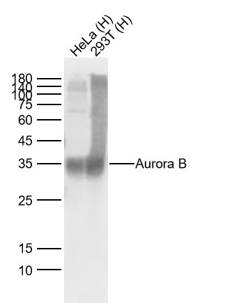 , ,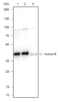 , ,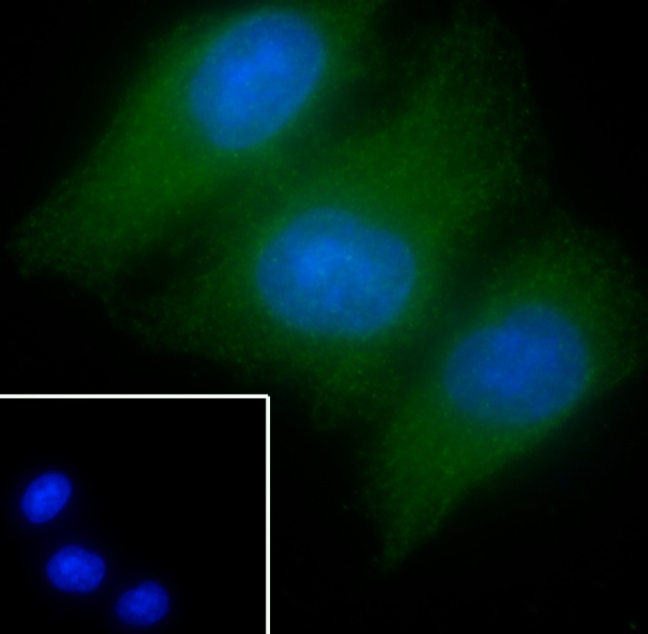 , ,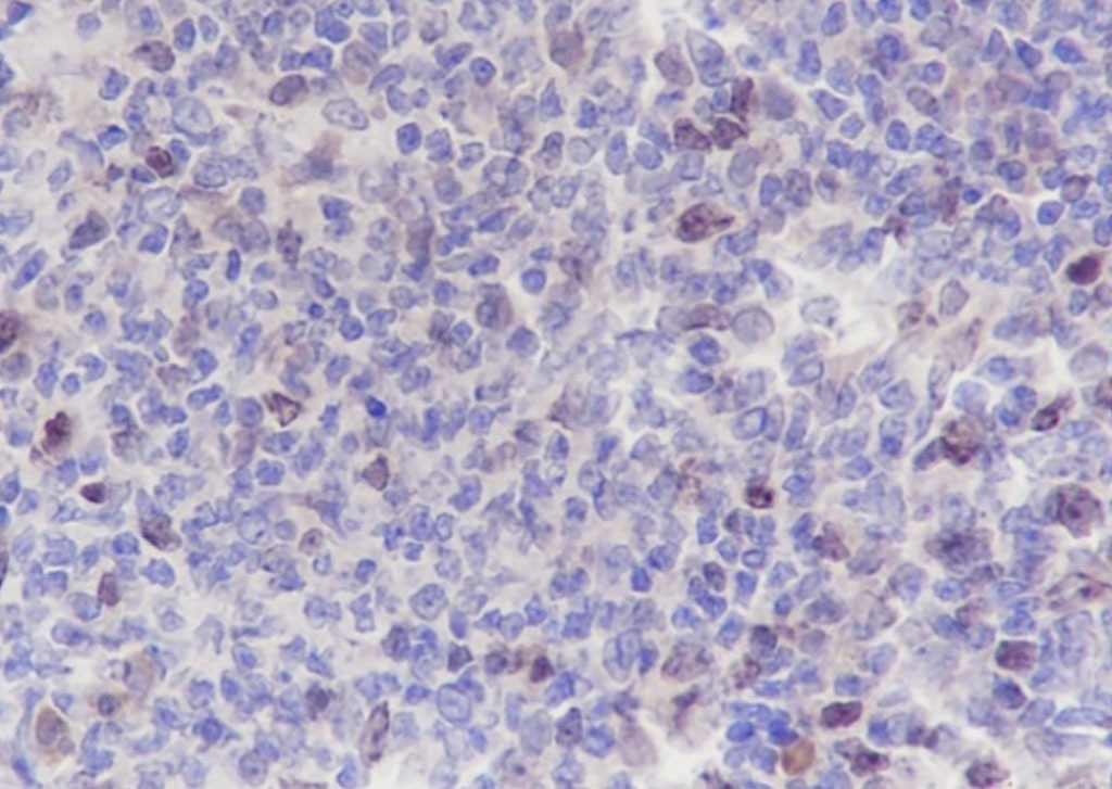 , ,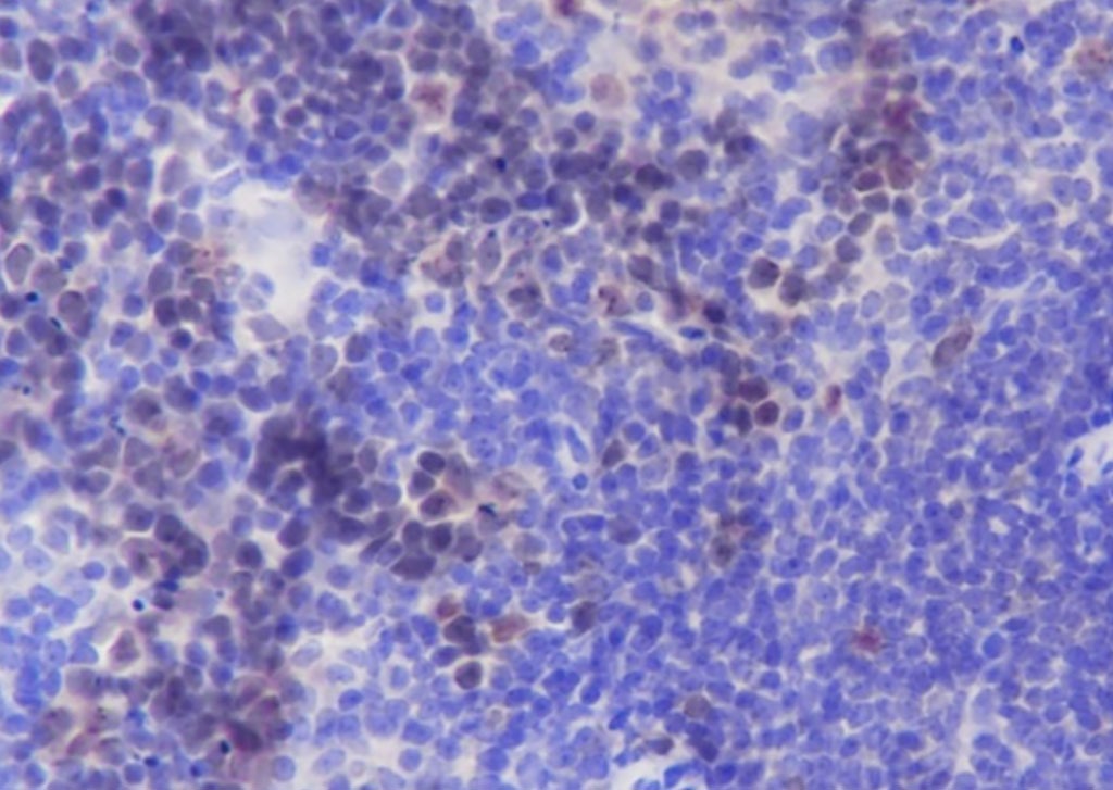 |
| Application | |
| Recommended Dose | ICC/IF=1:50-200; IF=1:50-200; IHC-Fr=1:50-200; IHC-P=1:50-200; WB=1:500-2000 |
| Antibody Type | Monoclonal |
| Host Species | Rabbit |
| Subcellular Localization | Nucleus. Chromosome. Chromosome, centromere. Cytoplasm, cytoskeleton, spindle. Note=Localizes on chromosome arms and inner centromeres from prophase through metaphase and then transferring to the spindle midzone and midbody from anaphase through cytokinesis. Colocalized with gamma tubulin in the mid-body. Proper localization of the active, Thr-232-phosphorylated form during metaphase may be dependent upon interaction with SPDYC. |
| Tissue Specificity | High level expression seen in the thymus. It is also expressed in the spleen, lung, testis, colon, placenta and fetal liver. Expressed during S and G2/M phase and expression is up-regulated in cancer cells during M phase. |
| Construction | Recombinant Antibody |
| Purification | Protein G purified |
| Appearance | Liquid |
| Formulation | 0.01M TBS (pH7.4) with 1% BSA, 0.02% Proclin300 and 50% Glycerol. |
| Concentration | 1 mg/mL |
| Research Background | This gene encodes a member of the aurora kinase subfamily of serine/threonine kinases. The genes encoding the other two members of this subfamily are located on chromsomes 19 and 20. These kinases participate in the regulation of segregation of chromosomes during mitosis and meiosis through association with microtubules. A pseudogene of this gene is located on chromosome 8. Multiple transcript variants encoding different isoforms have been found for this gene. [provided by RefSeq, Feb 2012]. |
| Immunogen | KLH conjugated synthetic peptide: human Aurora B |
| Antigen Species | Human |
| Gene Name | AURKB |
| Gene ID | |
| Protein Name | Aurora kinase B |
| Uniprot ID | |
| Biology Area | Aurora,Aurora,Phosphorylation,Aurora |
| Function | Serine/threonine-protein kinase component of the chromosomal passenger complex (CPC), a complex that acts as a key regulator of mitosis. The CPC complex has essential functions at the centromere in ensuring correct chromosome alignment and segregation and is required for chromatin-induced microtubule stabilization and spindle assembly. Involved in the bipolar attachment of spindle microtubules to kinetochores and is a key regulator for the onset of cytokinesis during mitosis. Required for central/midzone spindle assembly and cleavage furrow formation. AURKB phosphorylates the CPC complex subunits BIRC5/survivin, CDCA8/borealin and INCENP. Phosphorylation of INCENP leads to increased AURKB activity. Other known AURKB substrates involved in centromeric functions and mitosis are CENPA, DES/desmin, GPAF, KIF2C, NSUN2, RACGAP1, SEPT1, VIM/vimentin, GSG2/Haspin, and histone H3. A positive feedback loop involving GSG2 and AURKB contributes to localization of CPC to centromeres. Phosphorylation of VIM controls vimentin filament segregation in cytokinetic process, whereas histone H3 is phosphorylated at 'Ser-10' and 'Ser-28' during mitosis. A positive feedback between GSG2 and AURKB contributes to CPC localization. AURKB is also required for kinetochore localization of BUB1 and SGOL1. Phosphorylation of p53/TP53 negatively regulates its transcriptional activity. |
| Molecular Weight | Theoretical: 39 kDa. Actual: 35 kDa. |
| Stability & Storage | Store at -20°C or -80°C for 12 months. Avoid repeated freeze-thaw cycles. |
| Transport | Shipping with blue ice. |
| Size | Quantity | Unit Price | Amount | Operation |
|---|
 Hello! How can I help you today?
 Copyright © 2015-2026 TargetMol Chemicals Inc. All Rights Reserved.
