Shopping Cart
Remove All Your shopping cart is currently empty
Anti-VE-Cadherin Polyclonal Antibody 2 is a Rabbit antibody targeting VE-Cadherin. Anti-VE-Cadherin Polyclonal Antibody 2 can be used in FCM, WB.
| Pack Size | Price | USA Warehouse | Global Warehouse | Quantity |
|---|---|---|---|---|
| 50 μL | $220 | 7-10 days | 7-10 days | |
| 100 μL | $373 | 7-10 days | 7-10 days | |
| 200 μL | $527 | 7-10 days | 7-10 days |
| Description | Anti-VE-Cadherin Polyclonal Antibody 2 is a Rabbit antibody targeting VE-Cadherin. Anti-VE-Cadherin Polyclonal Antibody 2 can be used in FCM, WB. |
| Synonyms | VECD, VE-Cad, VEcad, Vec, Cd144, cadherin 5, type 2 (vascular endothelium), AA408225, 7B4 |
| Ig Type | IgG |
| Reactivity | Human (predicted:Mouse,Rat) |
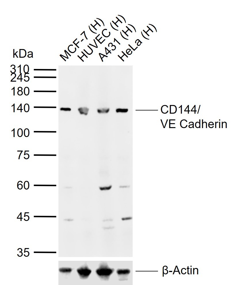
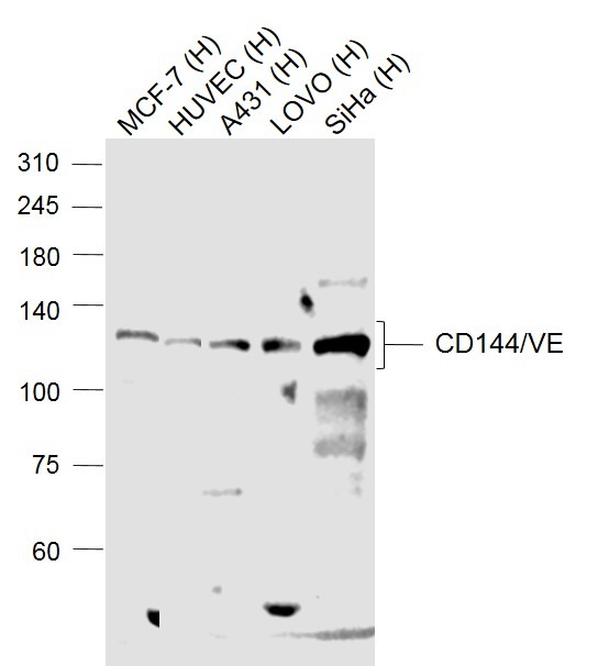
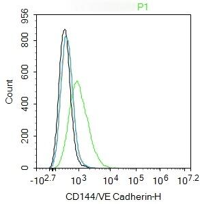
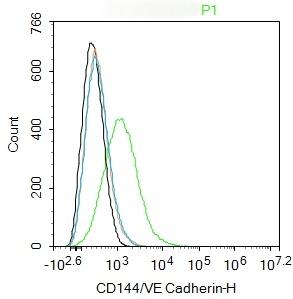
| Verified Activity | 1. Sample: Lane 1: Human MCF-7 cell lysates Lane 2: Human HUVEC cell lysates Lane 3: Human A431 cell lysates Lane 4: Human HeLa cell lysates Primary: Anti-CD144/VE Cadherin (TMAB-01946) at 1/1000 dilution Secondary: IRDye800CW Goat Anti-Rabbit IgG at 1/20000 dilution Predicted band size: 86 kDa Observed band size: 140 kDa 2. Sample: Lane 1: MCF-7 (Human) Cell Lysate at 30 μg Lane 2: HUVEC (Human) Cell Lysate at 30 μg Lane 3: A431 (Human) Cell Lysate at 30 μg Lane 4: LOVO (Human) Cell Lysate at 30 μg Lane 5: SiHa (Human) Cell Lysate at 30 μg Primary: Anti-CD144/VE Cadherin (TMAB-01946) at 1/1000 dilution Secondary: IRDye800CW Goat Anti-Rabbit IgG at 1/20000 dilution Predicted band size: 130 kDa Observed band size: 130 kDa 3. Blank control: HUVEC. Primary Antibody (green line): Rabbit Anti-CD144/VE Cadherin antibody (TMAB-01946) Dilution: 1 μg/Test; Secondary Antibody (white blue line): Goat anti-rabbit IgG-AF488 Dilution: 0.5 μg/Test. Isotype control (orange line): Normal Rabbit IgG Protocol The cells were incubated in 5% BSA to block non-specific protein-protein interactions for 30 min at room temperature. Cells stained with Primary Antibody for 30 min at room temperature. The secondary antibody used for 40 min at room temperature. 4. Blank control: HUVC. Primary Antibody (green line): Rabbit Anti-CD144/VE Cadherin antibody (TMAB-01946) Dilution: 1 μg/Test; Secondary Antibody (white blue line): Goat anti-rabbit IgG-AF488 Dilution: 0.5 μg/Test. Isotype control (orange line): Normal Rabbit IgG Protocol The cells were incubated in 5% BSA to block non-specific protein-protein interactions for 30 min at room temperature. Cells stained with Primary Antibody for 30 min at room temperature. The secondary antibody used for 40 min at room temperature.     |
| Application | |
| Recommended Dose | WB: 1:500-2000; FCM: 1ug/Test |
| Antibody Type | Polyclonal |
| Host Species | Rabbit |
| Subcellular Localization | Cell junction. Cell membrane. Found at cell-cell boundaries and probably at cell-matrix boundaries. |
| Tissue Specificity | Endothelial tissues and brain. |
| Construction | Polyclonal Antibody |
| Purification | Protein A purified |
| Appearance | Liquid |
| Formulation | 0.01M TBS (pH7.4) with 1% BSA, 0.02% Proclin300 and 50% Glycerol. |
| Concentration | 1 mg/mL |
| Research Background | This gene is a classical cadherin from the cadherin superfamily and is located in a six-cadherin cluster in a region on the long arm of chromosome 16 that is involved in loss of heterozygosity events in breast and prostate cancer. The encoded protein is a calcium-dependent cell-cell adhesion glycoprotein comprised of five extracellular cadherin repeats, a transmembrane region and a highly conserved cytoplasmic tail. Functioning as a classic cadherin by imparting to cells the ability to adhere in a homophilic manner, the protein may play an important role in endothelial cell biology through control of the cohesion and organization of the intercellular junctions. An alternative splice variant has been described but its full length sequence has not been determined. [provided by RefSeq, Jul 2008]. |
| Immunogen | KLH conjugated synthetic peptide: mouse CD144/VE Cadherin |
| Antigen Species | Mouse |
| Gene Name | CDH5 |
| Gene ID | |
| Protein Name | Cadherin-5 |
| Uniprot ID | |
| Biology Area | Cadherins,Endothelial Cell Markers,Cell adhesion molecules,Endothelial Cells,Endothelium,Cadherins,Endothelial,Vascular |
| Function | Cadherins are calcium dependent cell adhesion proteins. They preferentially interact with themselves in a homophilic manner in connecting cells; cadherins may thus contribute to the sorting of heterogeneous cell types. This cadherin may play a important role in endothelial cell biology through control of the cohesion and organization of the intercellular junctions. It associates with alpha-catenin forming a link to the cytoskeleton. Acts in concert with KRIT1 to establish and maintain correct endothelial cell polarity and vascular lumen. These effects are mediated by recruitment and activation of the Par polarity complex and RAP1B. Required for activation of PRKCZ and for the localization of phosphorylated PRKCZ, PARD3, TIAM1 and RAP1B to the cell junction. |
| Molecular Weight | Theoretical: 86 kDa. |
| Stability & Storage | Store at -20°C or -80°C for 12 months. Avoid repeated freeze-thaw cycles. |
| Transport | Shipping with blue ice. |
| Size | Quantity | Unit Price | Amount | Operation |
|---|

Copyright © 2015-2026 TargetMol Chemicals Inc. All Rights Reserved.