
Shopping Cart
Remove All Your shopping cart is currently empty

Synonyms: Tie-2, Tie2, TEK tyrosine kinase, endothelial, STK1, Hyk, Cd202b, AA517024
Anti-TIE2 Polyclonal Antibody
| Pack Size | Price | USA Stock | Global Stock | Quantity |
|---|---|---|---|---|
| 50 µL | $220 | 7-10 days | 7-10 days | |
| 100 µL | $374 | 7-10 days | 7-10 days | |
| 200 µL | $527 | 7-10 days | 7-10 days |
| Description | Anti-TIE2 Polyclonal Antibody is a Rabbit antibody targeting TIE2. Anti-TIE2 Polyclonal Antibody can be used in FCM, ICC/IF. |
| Synonyms | Tie-2, Tie2, TEK tyrosine kinase, endothelial, STK1, Hyk, Cd202b, AA517024 |
| Ig Type | IgG |
| Reactivity | Human (predicted:Mouse,Rat,Pig,Cow,Horse) |
| Verified Activity | 1. Tissue/cell: A549 cell; 4% Paraformaldehyde-fixed; Triton X-100 at room temperature for 20 min; Blocking buffer (normal goat serum) at 37°C for 20 min; Antibody incubation with (TIE2) polyclonal Antibody, Unconjugated (TMAB-01839) 1:100, 90 minutes at 37°C; followed by a FITC conjugated Goat Anti-Rabbit IgG antibody at 37°C for 90 minutes, DAPI (blue) was used to stain the cell nucleus. 2. Blank control (Black line): HUVEC (Black). Primary Antibody (green line): Rabbit Anti-TIE2 antibody (TMAB-01839) Dilution: 1 μg/10^6 cells; Isotype Control Antibody (orange line): Rabbit IgG. Secondary Antibody (white blue line): Goat anti-rabbit IgG-AF647 Dilution: 1 μg/test. Protocol The cells were fixed with 4% PFA (10 min at room temperature) and then permeabilized with 0.1% PBST for 20 min at room temperature. The cells were then incubated in 5% BSA to block non-specific protein-protein interactions for 30 min at room temperature. Cells stained with Primary Antibody for 30 min at room temperature. The secondary antibody used for 40 min at room temperature. 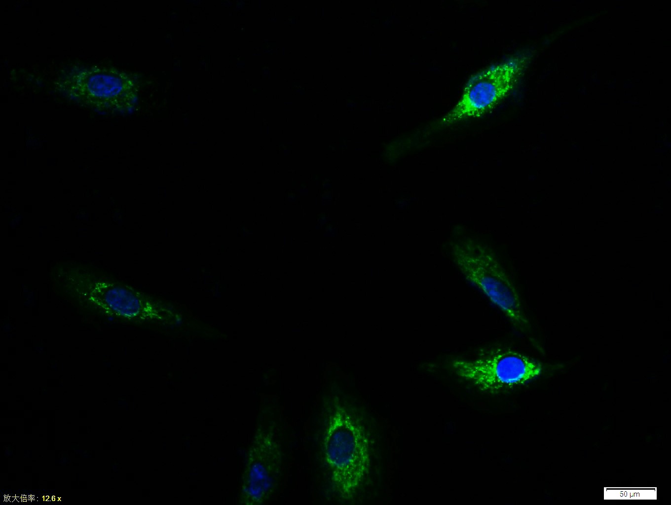 , ,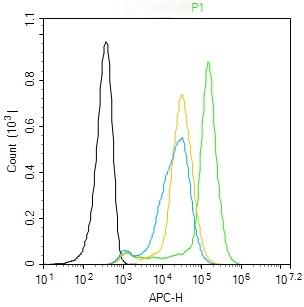 |
| Application | |
| Recommended Dose | FCM=3 μg/Test; ICC/IF=1:100-500 |
| Antibody Type | Polyclonal |
| Host Species | Rabbit |
| Subcellular Localization | Cell membrane; Single-pass type I membrane protein. Cell junction. Cell junction, focal adhesion. Cytoplasm, cytoskeleton. Secreted. Note=Recruited to cell-cell contacts in quiescent endothelial cells. Colocalizes with the actin cytoskeleton and at actin stress fibers during cell spreading. Recruited to the lower surface of migrating cells, especially the rear end of the cell. Proteolytic processing gives rise to a soluble extracellular domain that is secreted. |
| Tissue Specificity | Predominantly expressed in endothelial cells and their progenitors, the angioblasts. Has been directly found in placenta and lung, with a lower level in umbilical vein endothelial cells, brain and kidney. |
| Construction | Polyclonal Antibody |
| Purification | Protein A purified |
| Appearance | Liquid |
| Formulation | 0.01M TBS (pH7.4) with 1% BSA, 0.02% Proclin300 and 50% Glycerol. |
| Concentration | 1 mg/mL |
| Research Background | The TEK receptor tyrosine kinase is expressed almost exclusively in endothelial cells in mice, rats, and humans. This receptor possesses a unique extracellular domain containing 2 immunoglobulin-like loops separated by 3 epidermal growth factor-like repeats that are connected to 3 fibronectin type III-like repeats. The ligand for the receptor is angiopoietin-1. Defects in TEK are associated with inherited venous malformations; the TEK signaling pathway appears to be critical for endothelial cell-smooth muscle cell communication in venous morphogenesis.TEK is closely related to the TIE receptor tyrosine kinase. |
| Immunogen | KLH conjugated synthetic peptide: human Tie2 |
| Antigen Species | Human |
| Gene Name | TEK |
| Gene ID | |
| Protein Name | Angiopoietin-1 receptor |
| Uniprot ID | |
| Biology Area | Endothelial Markers,Receptor Tyrosine Kinases,Angiogenesis and vasculogenesis,Endothelium,Endothelial Cells,Endothelial Cell Markers,Growth factor receptors |
| Function | This protein is a protein tyrosine-kinase transmembrane receptor for angiopoietin 1. It may constitute the earliest mammalian endothelial cell lineage marker. Probably regulates endothelial cell proliferation, differentiation and guides the proper patterning of endothelial cells during blood vessel formation. |
| Molecular Weight | Theoretical: 124 kDa. |
| Stability & Storage | Store at -20°C or -80°C for 12 months. Avoid repeated freeze-thaw cycles. |
| Transport | Shipping with blue ice. |
| Size | Quantity | Unit Price | Amount | Operation |
|---|
 Hello! How can I help you today?
 Copyright © 2015-2026 TargetMol Chemicals Inc. All Rights Reserved.
