
Shopping Cart
Remove All Your shopping cart is currently empty

Synonyms:
Anti-PODXL Polyclonal Antibody
| Pack Size | Price | USA Stock | Global Stock | Quantity |
|---|---|---|---|---|
| 50 µL | $221 | 7-10 days | 7-10 days | |
| 100 µL | $372 | 7-10 days | 7-10 days | |
| 200 µL | $527 | 7-10 days | 7-10 days |
| Description | Anti-PODXL Polyclonal Antibody is a Rabbit antibody targeting PODXL. Anti-PODXL Polyclonal Antibody can be used in FCM,IF,IHC-Fr,IHC-P,WB. |
| Ig Type | IgG |
| Reactivity | Human,Mouse,Rat |
| Verified Activity | 1. Tissue/cell: human kidney tissue; 4% Paraformaldehyde-fixed and paraffin-embedded; Antigen retrieval: citrate buffer (0.01M, pH6.0), Boiling bathing for 15 min; Block endogenous peroxidase by 3% Hydrogen peroxide for 30 min; Blocking buffer (normal goat serum) at 37°C for 20 min; Incubation: Anti-PCX Polyclonal Antibody, Unconjugated (TMAB-01558) 1:200, overnight at 4°C, followed by conjugation to the secondary antibody and DAb staining. 2. Sample: Kidney (Mouse) lysate at 30 μg; Primary: Anti-PODXL (TMAB-01558) at 1:300 dilution; Secondary: HRP conjugated Goat-Anti-Rabbit Igg (bse-0295G-HRP) at 1: 5000 dilution; ECL excitated the fluorescence; Predicted band size: 59 kDa Observed band size:59 kDa 3. Blank control: RSC96 (blue). Primary Antibody: Rabbit Anti-PODXL antibody ,used under the same conditions); Secondary Antibody: Goat anti-rabbit IgG-Pe (white blue), Dilution: 1:200 in 1 X PBS containing 0.5% BSA. Protocol The cells were fixed with 2% paraformaldehyde (10 min). Antibody (TMAB-01558, 0.2 μg/1x10^6 cells) were incubated for 30 min on the ice, followed by 1 X PBS containing 0.5% BSA + 10% goat serum (15 min) to block non-specific protein-protein interactions. Then the Goat Anti-rabbit IgG/PE antibody was added into the blocking buffer mentioned above to react with the primary antibody of TMAB-01558 at 1/200 dilution for 30 min on ice. 4. Sample: Liver (Mouse) Lysate at 40 μg Primary: Anti-PODXL (TMAB-01558) at 1/1000 dilution Secondary: IRDye800CW Goat Anti-Rabbit IgG at 1/20000 dilution Predicted band size: 59 kDa Observed band size: 59 kDa 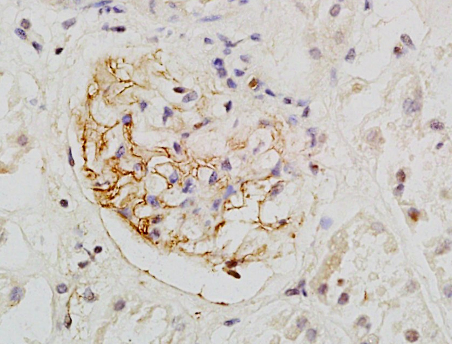 , ,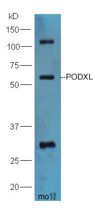 , ,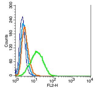 , ,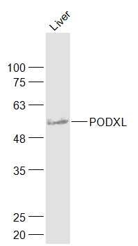 |
| Application | |
| Recommended Dose | WB: 1:500-2000; IHC-P: 1:100-500; IHC-Fr: 1:100-500; IF: 1:100-500; FCM: 1μg/Test |
| Antibody Type | Polyclonal |
| Host Species | Rabbit |
| Subcellular Localization | Apical cell membrane. Cell projection, lamellipodium. Cell projection, filopodium. Cell projection, ruffle. Cell projection, microvillus. Membrane raft. Membrane. In single attached epithelial cells is restricted to a preapical pole on the free plasma membrane whereas other apical and basolateral proteins are not yet polarized. Colocalizes with SLC9A3R2 at the apical plasma membrane during epithelial polarization. Colocalizes with SLC9A3R1 at the trans-Golgi network (transiently) and at the apical plasma membrane. Its association with the membrane raft is transient. Colocalizes with actin filaments, EZR and SLC9A3R1 in a punctate pattern at the apical cell surface where microvilli form. Colocalizes with EZR and SLC9A3R2 at the apical cell membrane of glomerular epithelium cells (By similarity). Forms granular, punctuated pattern, forming patches, preferentially adopting a polar distribution, located on the migrating poles of the cell or forming clusters along the terminal ends of filipodia establishing contact with the endothelial cells. Colocalizes with the submembrane actin of lamellipodia, particularly associated with ruffles. Colocalizes with vinculin at protrusions of cells. Colocalizes with ITGB1. Colocalizes with PARD3, PRKCI, EXOC5, OCLN, RAB11A and RAB8A in apical membrane initiation sites (AMIS) during the generation of apical surface and luminogenesis. |
| Tissue Specificity | Glomerular epithelium cell (podocyte). |
| Construction | Polyclonal Antibody |
| Purification | Protein A purified |
| Appearance | Liquid |
| Formulation | 0.01M TBS (pH7.4) with 1% BSA, 0.02% Proclin300 and 50% Glycerol. |
| Concentration | 1 mg/mL |
| Research Background | This gene encodes a member of the sialomucin protein family. The encoded protein was originally identified as an important component of glomerular podocytes. Podocytes are highly differentiated epithelial cells with interdigitating foot processes covering the outer aspect of the glomerular basement membrane. Other biological activities of the encoded protein include: binding in a membrane protein complex with Na+/H+ exchanger regulatory factor to intracellular cytoskeletal elements, playing a role in hematopoetic cell differentiation, and being expressed in vascular endothelium cells and binding to L-selectin. [provided by RefSeq, Jul 2008] |
| Immunogen | KLH conjugated synthetic peptide: human PODXL |
| Antigen Species | Human |
| Gene Name | PODXL |
| Gene ID | |
| Protein Name | Podocalyxin |
| Uniprot ID | |
| Biology Area | Tumor biomarkers,Endothelial Cell Markers,Endothelium,Focal Adhesions,Surface Molecules |
| Function | Involved in the regulation of both adhesion and cell morphology and cancer progression. Function as an anti-adhesive molecule that maintains an open filtration pathway between neighboring foot processes in the podocyte by charge repulsion. Acts as a pro-adhesive molecule, enhancing the adherence of cells to immobilized ligands, increasing the rate of migration and cell-cell contacts in an integrin-dependent manner. Induces the formation of apical actin-dependent microvilli. Involved in the formation of a preapical plasma membrane subdomain to set up inital epithelial polarization and the apical lumen formation during renal tubulogenesis. Plays a role in cancer development and aggressiveness by inducing cell migration and invasion through its interaction with the actin-binding protein EZR. Affects EZR-dependent signaling events, leading to increased activities of the MAPK and PI3K pathways in cancer cells. |
| Molecular Weight | Theoretical: 59 kDa. Actual: 59 kDa. |
| Stability & Storage | Store at -20°C or -80°C for 12 months. Avoid repeated freeze-thaw cycles. |
| Transport | Shipping with blue ice. |
| Size | Quantity | Unit Price | Amount | Operation |
|---|
 Hello! How can I help you today?
 Copyright © 2015-2026 TargetMol Chemicals Inc. All Rights Reserved.
