
Shopping Cart
Remove All Your shopping cart is currently empty


Synonyms:
| Pack Size | Price | USA Stock | Global Stock | Quantity |
|---|---|---|---|---|
| 10 mg | $30 | In Stock | In Stock | |
| 25 mg | $48 | In Stock | In Stock | |
| 50 mg | $71 | In Stock | In Stock | |
| 100 mg | $106 | In Stock | In Stock |
| Description | 2-Aminoacridone is a widely used fluorophore (λexc=428 nm, λem=525 nm). |
| In vitro | By using 2-Aminoacridone as labeling molecule, sensitivity for the detection of GAG-derived disaccharides is greatly enhanced, and resolution is also improved. |
| Cell Research | Instructions for use I. Preparation of solutions 1. Preparation of mother solution and working solution: The concentration of 2-Aminoacridone is usually between 1–10 μM in fluorescence experiments, and the specific concentration is adjusted according to the experimental requirements. For more sensitive applications, the concentration may be lower (e.g. 0.1 μM), while for experiments with larger sample volumes or requiring strong signals, the concentration can be appropriately increased (e.g. 10 μM). 2. Preparation of working solution: When performing fluorescence quantitative analysis, the amount of 2-Aminoacridone used depends on the concentration of the target molecule and the sensitivity requirements of the experiment. Generally, the concentration of the labeled solution should match the concentration of the substance to be tested to ensure optimal signal intensity. I. As a fluorescent probe Method: Dissolve 2-Aminoacridone in an appropriate solvent (such as DMSO, methanol or water). Before use, incubate it with the DNA, RNA or target molecule in the sample, usually for a short incubation time (1–2 hours). The labeling can then be analyzed using a fluorescence microscope or other fluorescence detection equipment. 2. Used for fluorescence quantitative analysis Method: In fluorescence analysis, 2-Aminoacridone can be used as a fluorescent marker to detect the DNA or RNA content in the sample. Common experimental methods include using a fluorescence spectrophotometer or a fluorescence microplate reader. The fluorescence intensity in the sample is proportional to its molecular number and is suitable for quantitative analysis. 3. Fluorescence staining: Method: In the fluorescence staining of cells or tissues, 2-Aminoacridone is used as a dye and directly added to the sample to stain the cells. After staining, a fluorescence microscope is used for imaging to observe the marker signal in the cell, with an excitation wavelength of 428 nm and an emission wavelength of 525 nm. The above information is based on published literature. Experimental procedures should be appropriately modified to meet specific research demands. |
| Molecular Weight | 210.23 |
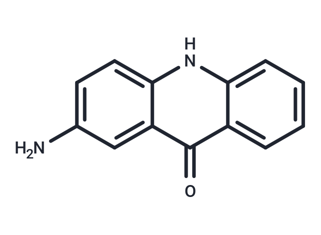
| Formula | C13H10N2O |
| Cas No. | 27918-14-5 |
| Smiles | Nc1ccc2[nH]c3ccccc3c(=O)c2c1 |
| Relative Density. | 1.306g/cm3 |
| Storage | Keep away from direct sunlight Powder: -20°C for 3 years Shipping with blue ice/Shipping at ambient temperature. | |||||||||||||||||||||||||||||||||||
| Solubility Information | DMSO: 60 mg/mL (285.4 mM), Sonication is recommended. | |||||||||||||||||||||||||||||||||||
Solution Preparation Table | ||||||||||||||||||||||||||||||||||||
DMSO
Note : The dilution table applies only to solid products. For liquid products, please calculate the stock solution based on the stated concentration and/or density. | ||||||||||||||||||||||||||||||||||||
 For example, if the intended dosage is 10 mg/kg for animals weighing 20 g , with a dosing volume of 100 μL per animal, and a total of 10 animals are to be administered, using a formulation of
  10% DMSO+ 40% PEG300+ 5% Tween 80+ 45% Saline/PBS/ddH2O , the resulting working solution concentration would be 2 mg/mL.
Dissolve 2 mg of the compound in 100 μL DMSO to obtain a stock solution at a concentration of 20 mg/mL . If the required concentration exceeds the compound's known solubility, please contact us for technical support before proceeding.
1) Add 100 μL of the DMSO stock solution to 400 µL PEG300 and mix thoroughly until the solution becomes clear.
2) Add 50 µL Tween 80 and mix well until fully clarified.
3) Add 450 µL Saline,PBS or ddH2O and mix thoroughly until a homogeneous solution is obtained.
| Size | Quantity | Unit Price | Amount | Operation |
|---|
 Hello! How can I help you today?
 Copyright © 2015-2026 TargetMol Chemicals Inc. All Rights Reserved.
