
Shopping Cart
Remove All Your shopping cart is currently empty


Synonyms:
| Pack Size | Price | USA Stock | Global Stock | Quantity |
|---|---|---|---|---|
| 1 mL | $61 | In Stock | In Stock |
| Hoechst 33342 Staining Solution | Specifications |
|---|---|
| Ingredient | Hoechst 33342 trihydrochloride |
| CAS | 875756-97-1 |
| Conc. | 5 mg/mL |
| Solvent | ddH2O |
1.Strong specificity in nuclear marker: with significantly higher affinity for DNA than RNA, resulting in minimal RNA background interference.
2.Low cytotoxicity: suitable for live-cell labeling studies.
3.Good compatibility: allowing co-staining with multiple fluorescent probes.
4.Simple operation and rapid detection.
Cell apoptosis detection;
Cell cycle analysis;
Cell viability assessment;
Drug toxicology evaluation.
Dilute the Hoechst 33342 stock solution with PBS to prepare a Hoechst 33342 staining working solution at a concentration of 0.5-10 μg/mL (protect from light). The specific concentration should be adjusted according to the sample and experimental requirements.
Staining of Adherent Cells
(1) Remove the old culture medium and wash the cells with an appropriate amount of PBS 1-3 times, 3 minutes each time.
(2) (Optional) Add 4% paraformaldehyde and fix the cells at room temperature for 10 minutes. Remove the fixative and wash the cells with PBS 3 times, 3 minutes each time.
(3) (Optional) If immunofluorescence staining is required, perform immunofluorescence staining first, followed by Hoechst 33342 staining. If no other staining is needed, proceed directly to Hoechst 33342 staining.
(4) Add an appropriate amount of Hoechst 33342 working solution to cover the cells. For fixed tissues or cells, incubate at room temperature in the dark for 3-8 minutes; for live cells or cultured tissues, incubate at 37 ℃ in the dark for 20-30 minutes.
(5) After incubation, remove the staining solution and wash the cells with PBS 2-3 times, 3 minutes each time.
(6) Add an appropriate amount of antifade mounting medium onto a microscope slide. Place the coverslip (with the cells facing downward) onto the medium, then seal the edges with mounting solution.
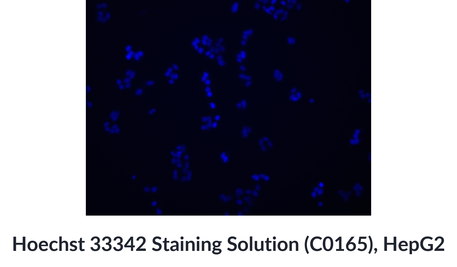
(7) Observe and capture images under a fluorescence microscope. Excitation wavelength (Ex) = 350 nm; Emission wavelength (Em) ≈ 461 nm.
Staining of Suspension Cells
(1) Collect suspension cells by centrifugation at 600 × g, 4 °C for 5 minutes, and discard the supernatant. Resuspend the cells in PBS and centrifuge again at 600 × g, 4 °C for 5 minutes. Discard the supernatant. Wash the cells with PBS 0–2 times.
(2) (Optional) Add 4% paraformaldehyde and fix at room temperature for 10 minutes. Remove the fixative and wash the cells three times with PBS, 3 minutes each time.
(3) (Optional) If immunofluorescence staining is required, perform immunofluorescence staining first, followed by Hoechst 33342 staining. If no other staining is needed, proceed directly to Hoechst 33342 staining.
(4) Resuspend the cells in an appropriate volume of Hoechst 33342 working solution. For fixed tissues or cells, incubate at room temperature in the dark for 3–8 minutes. For live cells or cultured tissues, incubate at 37 °C in the dark for 20-30 minutes. After incubation, remove the staining solution.
(5) Resuspend the cells in PBS, centrifuge at 600 × g, 4 °C for 5 minutes, and discard the supernatant. Repeat this step 1-2 times.
(6) Resuspend the cells in PBS for flow cytometry analysis or prepare cell smears for observation under a fluorescence microscope. Excitation wavelength (Ex) = 350 nm; Emission wavelength (Em) ≈ 461 nm.
Store at -20 ℃, protected from light for 12 months.
1.Hoechst 33342 is a fluorescent dye that is prone to photobleaching, so it should be handled protected from light. Experimental results should be detected as soon as possible after staining.
2.Since staining efficiency can be affected by sample type and experimental conditions, it is recommended to optimize the working concentration and staining time of Hoechst 33342 through preliminary experiments.
3.The product is for R&D use only, not for diagnostic procedures, food, drug, household or other uses.
4.Please wear a lab coat and disposable gloves.
| Size | Quantity | Unit Price | Amount | Operation |
|---|
 Hello! How can I help you today?
 Copyright © 2015-2026 TargetMol Chemicals Inc. All Rights Reserved.
