 Your shopping cart is currently empty


Drug Affinity Responsive Target Stability (DARTS) Assay Kit
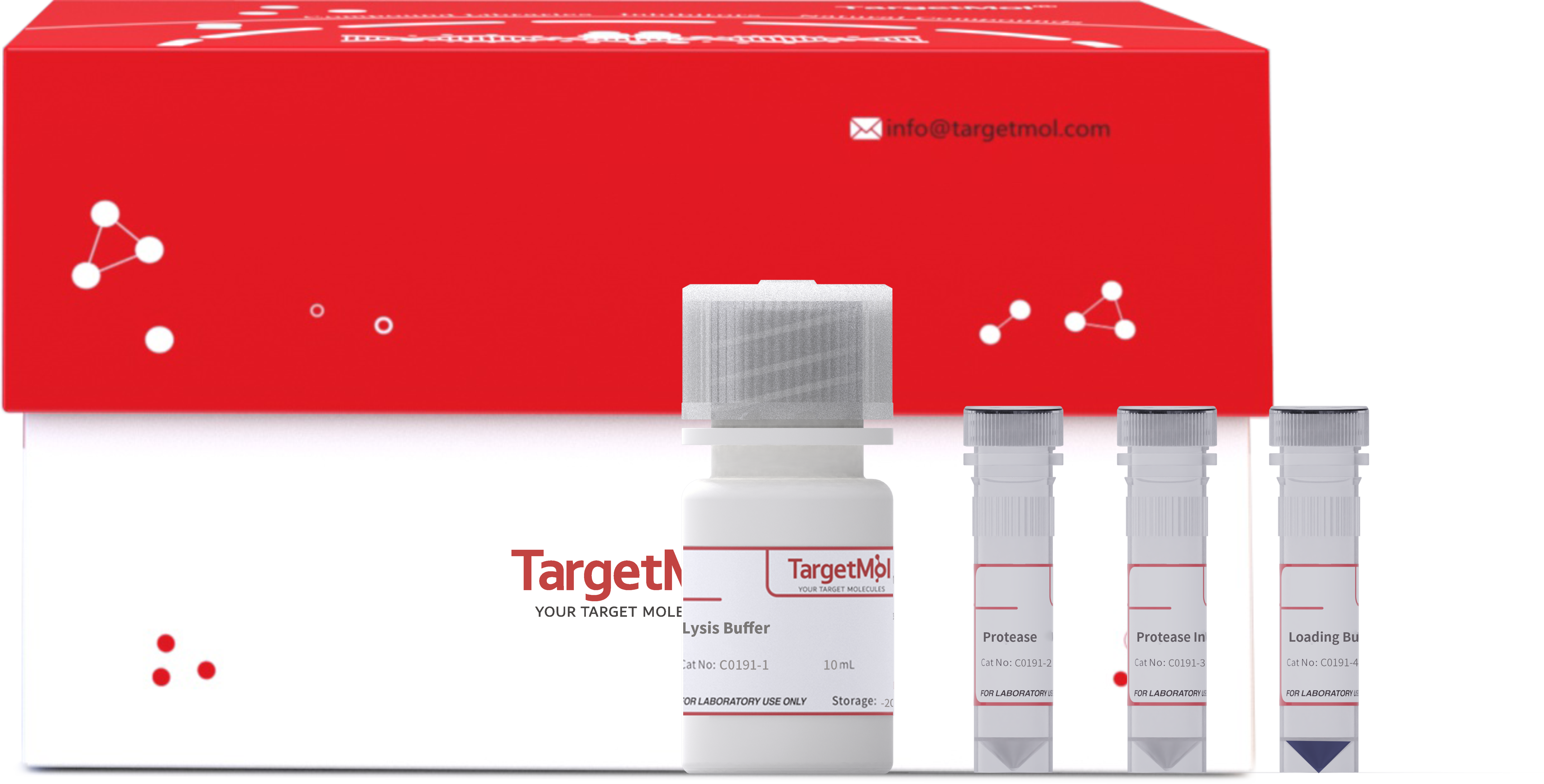
This kit provides the key buffers and protease digestion systems required for DARTS experiments, enabling efficient and sensitive detection of compound–target protein interactions. The method requires no compound labeling or modification, features a simple workflow and intuitive results, and can be combined with Western blotting, LC-MS/MS, or proteomics analysis. It offers an efficient and reliable experimental solution for drug discovery, target identification, and mechanism research.
| Pack Size | Price | USA Warehouse | Global Warehouse | Quantity |
|---|---|---|---|---|
| 100 T | $226 | - | In Stock | |
| 100 T * 5 | $678 | - | In Stock |
 Principle of Product
The basic principle of DARTS is that when a small-molecule compound binds to its target protein, the protein undergoes a conformational change in its three-dimensional structure, reducing its sensitivity to proteases (such as trypsin or thermolysin) and thereby exhibiting increased ”structural stability”.
In samples without the compound, the protein is easily hydrolyzed and degraded by proteases; in contrast, in the group treated with small-molecule drugs, the target protein bound to the drug is protected from degradation. By comparing the differences in protein remaining between the two groups, one can preliminarily determine whether the drug binds to the protein.
The core advantages of the DARTS method are:
-
No chemical modification or labeling of the drug is required, avoiding changes to the drug’s physicochemical properties or biological activity;
-
Applicable to complex natural systems (such as cell lysates or tissue extracts), reflecting drug–protein interactions under conditions close to the physiological environment;
-
Can be combined with Western blot, SDS-PAGE, silver staining, Coomassie staining, or mass spectrometry (LC-MS/MS) to achieve a complete workflow from qualitative validation to target identification.
 Product Information
E.g., Taking 100 T packing for example
| Catalog No. | Product Name | Packing |
|---|---|---|
| C0191-1 | Lysis Buffer | 10 mL |
| C0191-2 | Protease | 2 mg |
| C0191-3 | Protease Inhibitor Cocktail (100×) | 0.1 mL |
| C0191-4 | Loading Buffer (5×) | 3 mL |
 Features
- Easy to use: Provides a complete buffer and protease system, no additional optimization required.
- High sensitivity: Capable of detecting structural stability differences caused by drug-protein interactions at the nanomolar level.
- Broad application: Suitable for cell lysates from various sources, tissue samples, or recombinant protein systems.
- Clear results: Protein degradation in drug-treated vs. control groups can be easily compared via Western blot or gel staining.
- Compatible with multiple downstream analyses: Can be combined with LC-MS/MS for whole-proteome screening to identify potential new targets.
 Application
Target screening and validation for small-molecule drugs, study of protein-ligand interactions, and investigation of drug mechanisms of action.

Figure 1. The DARTS Assay Kit was used to assess the effect of Acevaltrate (ACE) on the stability of the PCBP1 protein.
After lysing HCT116 cells (human colorectal cancer cells), 5 µL of ACE solutions at concentrations of 0, 0.125, 0.25, 0.5, 1, and 2 mM were added to each 100 µL sample containing 100 µg total protein, resulting in final drug concentrations of 0, 6.25, 12.5, 25, 50, and 100 µM. The mixtures were incubated on a rotary shaker at room temperature for 60 min. Next, 4 µL of diluted protease (0.1 µg/µL) (Protease:Protein = 1:250) was added, mixed, and incubated at 37ºC for 30 min. The reaction was then stopped by adding 1 µL of Protease Inhibitor Cocktail (100×), followed by the addition of an appropriate amount of 5× loading buffer. Samples were denatured at 100ºC for 10 min. Finally, the digestion of PCBP1 protein was analyzed by Western blot. The results showed that ACE at 100 µM enhanced the stability of PCBP1 protein, suggesting that ACE may bind to PCBP1 and increase its resistance to proteolysis.(Actual results may vary depending on experimental conditions and detection instruments; the figure is for reference only).
Reference: Yu D, Hu H, Zhang Q, et al. Acevaltrate as a novel ferroptosis inducer with dual targets of PCBP1/2 and GPX4 in colorectal cancer. Signal Transduct Target Ther. 2025 Jul 7;10(1):211.
 Instructions
1.Cell Collection and Lysis:
After dispersing HCT116 cells, seed them into 100 mm culture dishes and culture until the cell density reaches 85%-90%. Aspirate the culture medium, wash the cells twice with PBS, and discard the PBS. Add 1 mL of PBS again, and quickly scrape the cells off the dish using a cell scraper (or collect them by trypsin digestion). Transfer the cells into a 1.5 mL centrifuge tube and centrifuge at 200 g for 5 min. Discard the PBS.
Add an appropriate amount of freshly prepared lysis buffer containing protease and phosphatase inhibitors, gently resuspend, and place on ice for 15 min to lyse the cells. Centrifuge at 12,000 rpm at 4°C for 15 min. Collect the supernatant and determine the protein concentration. BCA or Bradford assay is recommended for protein quantification. Adjust the protein concentration according to the measurement results to ensure it is within 1-5 mg/mL.
Note: Once the protein samples are prepared, it is recommended to proceed with subsequent experiments immediately. If storage is required, aliquot the samples into appropriate volumes and store at -80 ℃. For long-term storage, avoid repeated freeze-thaw cycles to prevent protein degradation or loss of activity.
2.Optimization of Digestion Ratios (It is recommended to confirm the digestion ratio first):
a) Prepare Protease Stock Solution: Dissolve in ultrapure water to a concentration of 10 µg/µL. Aliquot and store at -20 ℃, avoiding repeated freeze-thaw cycles. It is recommended to use within 6 months.
b) Preparation of enzyme working solutions at different ratios:
Take one tube of the stock solution and thaw it on ice. After thawing, add 4 µL of the stock to 196 µL of ultrapure water (diluting 50-fold, i.e., to 0.2 µg/µL). Prepare fresh before use, and adjust the dilution ratio as needed based on experimental results.
| Enzyme working solution ID | Enzyme-to-protein ratio | Total enzyme required (µg) | Enzyme concentration required (µg/µL) | Enzyme volume (µL) | Ultrapure water volume (µL) |
|---|---|---|---|---|---|
| A | 1:125 | 0.8 | 0.2 | 4 µL Enzyme Solution | 196 |
| B | 1:250 | 0.4 | 0.1 | 100 µL A | 100 |
| C | 1:500 | 0.2 | 0.05 | 100 µL B | 100 |
| D | 1:1000 | 0.1 | 0.025 | 100 µL C | 100 |
| E | 1:2000 | 0.05 | 0.0125 | 100 µL D | 100 |
| F | 1:4000 | 0.025 | 0.00625 | 100 µL E | 100 |
| G | 1:8000 | 0.0125 | 0.003125 | 100 µL F | 100 |
| H | 1:16000 | 0.00625 | 0.0015625 | 100 µL G | 100 |
- In the table, a total protein amount of 100 µg per sample is as an example. For a 2-fold dilution, prepare a total of 8 tubes of enzyme working solution. After preparation, tubes A–G contain 100 µL each, and tube H contains 200 µL. For the 8 protein sample tubes, add 4 µL of the enzyme working solution to each. Additionally, prepare one tube without enzyme, adding 4 µL of ultrapure water as a blank control.
c) After mixing each of the 9 samples, vortex and centrifuge briefly, then incubate at 37 ℃ for 30 min.
d) At the end of the incubation, immediately add Protease Inhibitor Cocktail (100x) to stop the digestion reaction.
e) Add an appropriate amount of Loading Buffer (5x) to each sample (for example, add 25 µL of Loading Buffer (5x) to a 100 µL reaction mixture), then heat at 100 ℃ for 10 min to terminate the reaction. Store the samples temporarily at -20 ℃.
f) Perform Western blot to detect the digestion of PCBP1 protein. The results are shown in the figure below.

Figure 2. Detection results of different protease-to-total protein ratios using DARTS Assay Kit.
For each group, 100 µL of HCT116 total protein (100 µg) was mixed with 4 µL of diluted protease working solution. After mixing, the samples were incubated at 37 ℃ for 30 min to allow protein digestion. At the end of incubation, 1 µL of Protease Inhibitor Cocktail (100x) was added to stop the reaction, followed by the addition of 5x loading buffer. The mixture was then heated at 100 ℃ for 10 min.
Western blot was subsequently used to assess the digestion of PCBP1 protein. The results showed that when the protease-to-protein ratio was approximately 1:250, significant changes in the protein bands were observed, facilitating subsequent experiments to determine whether small-molecule drugs affect protein stability.
- Actual results may vary depending on experimental conditions, detection instruments, and other factors; the results shown are for reference only.
3.Drug Treatment:
a) According to references or experimental design, prepare an appropriate concentration gradient of the drug and dilute stepwise from the stock solution to obtain the desired working concentrations. For example, concentrations can be set at 0, 0.125, 0.25, 0.5, 1, 2 mM, etc.
Each experiment should include two types of control groups: Negative Control: Protease is not added. Blank Control: Neither Protease nor drug is added; only the solvent corresponding to the drug (e.g., pure water or DMSO) is added.
It is recommended to add 100 µL of protein sample at 1 mg/mL per tube (i.e., total protein ~100 µg). The protein concentration and volume should be consistent with step 2b.
b) Add 5 µL of the drug working solution at the desired concentrations to each experimental tube. For the blank control group, add an equal volume of the drug solvent (e.g., pure water or DMSO). Gently mix and incubate on a shaker at room temperature for 60 min, or incubate in the dark at 4 ℃ overnight. The incubation temperature and duration can be adjusted according to the properties of the drug and reference protocols.
4.Enzymatic Digestion:
a) Preparation of Protease Stock Solution: Dissolve protease in ultrapure water to a final concentration of 10 µg/µL. Aliquot and store at -20 ℃ to avoid repeated freeze-thaw cycles. It is recommended to use within 6 months.
b) Preparation of Protease Working Solution: According to the protease-to-total protein ratio determined in Step 2, dilute the protease stock solution to prepare the desired concentration of the protease working solution. Perform the dilution on ice to maintain enzyme stability and activity.
c) Enzyme Reaction:
Add 4 µL of the pre-diluted protease working solution to each experimental group. For the negative control group, add an equal volume of ultrapure water instead of protease.
Note: Protein samples should always be kept on ice or at 4 ℃ before enzymatic digestion.
d) Enzymatic Incubation:
Gently mix and briefly centrifuge to collect the liquid at the bottom of the tube. Incubate the samples at 37 ℃ for 30 min to allow protease digestion.
e) Reaction Termination:
After incubation, immediately add Protease Inhibitor Cocktail (100x) to stop the enzymatic reaction (e.g., add 1 µL of Protease Inhibitor Cocktail (100x) to a 100 µL reaction system).
5.Sample Processing and Storage
The effects of a drug on target protein stability can be evaluated using various analytical techniques:
-
If the target protein is known, Western blot analysis can be used to assess changes in protease digestion resistance before and after drug treatment.
-
If the target protein is unknown, LC-MS/MS can be applied to identify proteins whose stability changes upon drug binding.
-
For target proteins with known enzymatic activity, enzyme activity assays can be used for high-throughput screening.
-
When the target protein is well characterized, ELISA or other detection methods can be selected as needed.
Example procedures for Western blot and LC-MS/MS analysis:
a) Add an appropriate volume of 5x loading buffer according to the sample volume (e.g., add 25 µL of 5x loading buffer to a 100 µL reaction system).
b) Heat the samples at 100°C for 10 minutes to ensure complete protein denaturation.
c) The heat-treated samples can be directly used for subsequent analyses. For validation of known drug targets, perform Western blot analysis; for the identification of unknown or novel target proteins, use LC-MS/MS or other proteomics approaches. If the samples are not to be used immediately, store at –80°C to prevent protein degradation or structural alterations.
 Storage
Store at -20 ℃ for one year.
 Precautions
1.Protein samples should be used as soon as possible after preparation to prevent protein degradation or loss of activity. Aliquot the samples and store at -80 °C. Avoid repeated freeze–thaw cycles.
2.Except for incubation and enzymatic digestion steps, all other operations (such as sample handling and enzyme dilution) should be performed on ice or at 4 °C to maintain protein stability and enzyme activity.
3.Protease concentration has a significant impact on experimental results. Excessive protease may cause nonspecific degradation. It is recommended to perform a preliminary experiment to determine the optimal digestion ratio.
4.Drug concentration, incubation time, and temperature should be optimized according to the specific properties of the compound or relevant literature. High drug concentrations or prolonged incubation may lead to nonspecific protein alterations.
5.Protease stock and working solutions should not be left standing for long periods. Prepare fresh solutions before use. After use, immediately seal the containers and store at –20 ℃.
6.After adding the Protease Inhibitor Cocktail (100×) to terminate the reaction, mix the solution immediately to ensure complete inhibition of enzymatic activity.
7.The product is for R&D use only, not for diagnostic procedures, food, drug, household or other uses.
8.Please wear a lab coat and disposable gloves.
 Instruction Manual
| Size | Quantity | Unit Price | Amount | Operation |
|---|

Copyright © 2015-2026 TargetMol Chemicals Inc. All Rights Reserved.


